
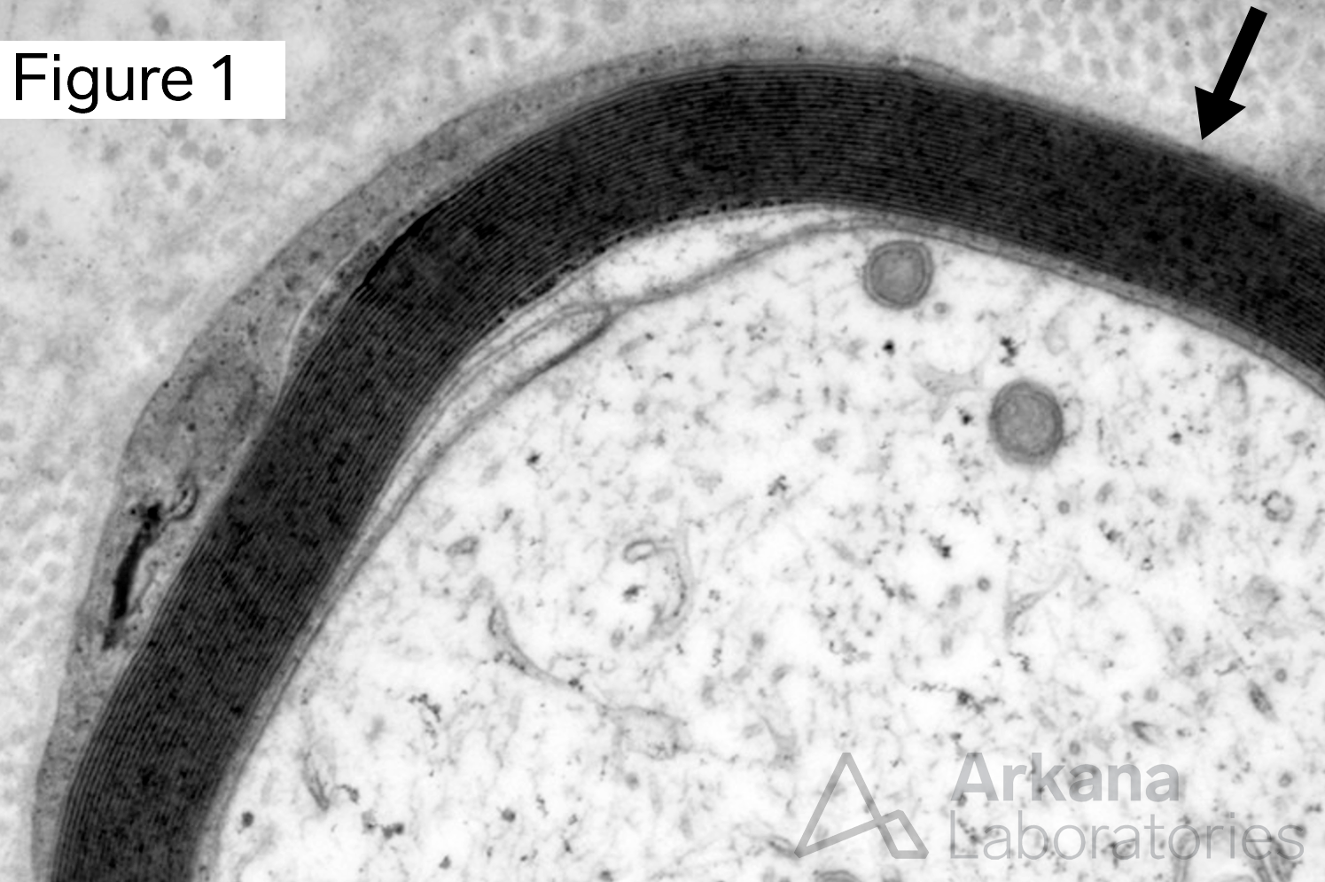
Figure 1 shows a myelin sheath (arrow) with orderly myelin wrapping. No abnormality of myelin periodicity is seen (i.e. no widely spaced or uncompact myelin is observed). No morphologic evidence to suggest a paraproteinopathy is identified. Original magnification: Electron microscopy, 20K.
A 55-year-old female patient presents with lower extremity weakness and numbness in her feet. Her medication list includes insulin. EMG/NCS showed electrodiagnostic evidence of denervation-type change. A biopsy of the sural nerve was performed. Neuropathologic review showed moderate to marked axonopathic changes characterized by relatively diffuse axonal loss, consistent with diabetic polyneuropathy. After reviewing Figure 1, which cell of the peripheral nervous system (PNS) is responsible for the orderly myelin sheath seen here by electron microscopy?
A. Schwann cell
B. Astrocyte
C. Oligodendrocyte
D. Microglia
Answer: Schwann cell
The Schwann cell is the myelinated cell of the peripheral nervous system (PNS), answer choice (A).
The oligodendrocyte (C) is the myelinating cell of the central nervous system (CNS).
Why were the other answers wrong?
Astrocytes (B) are glial cells of the CNS that have many functions, including regulating the blood brain barrier.
Microglia (D) are part of the immune defense of the CNS.
References
Brat DJ. Overview of central nervous system anatomy and histology. In: Neuropathology: A volume in the series: Foundations in diagnostic pathology, 2nd ed., Edited by RA Prayson. Elsevier Saunders, 2012: 1-39.
Quick note: This post is to be used for informational purposes only and does not constitute medical or health advice. Each person should consult their own doctor with respect to matters referenced. Arkana Laboratories assumes no liability for actions taken in reliance upon the information contained herein.