Clinical History
- This 60-year-old diabetic patient presented with complaints of bilateral chronic numbness and tingling involving their legs in a stock-type distribution. Electrodiagnostic studies showed features of a predominantly axonal distal sensorimotor neuropathy.
Which of the following do the small groups of large diameter myelinated axons represent?
These are best demonstrated on toluidine blue stained thick section and electron microscopy, but are also visible on H&E, modified Gomori Trichrome and neurofilament immunohistochemical stain.
A. Regenerated clusters
B. Active axon degeneration
C. Demyelination
D. Normal nerve
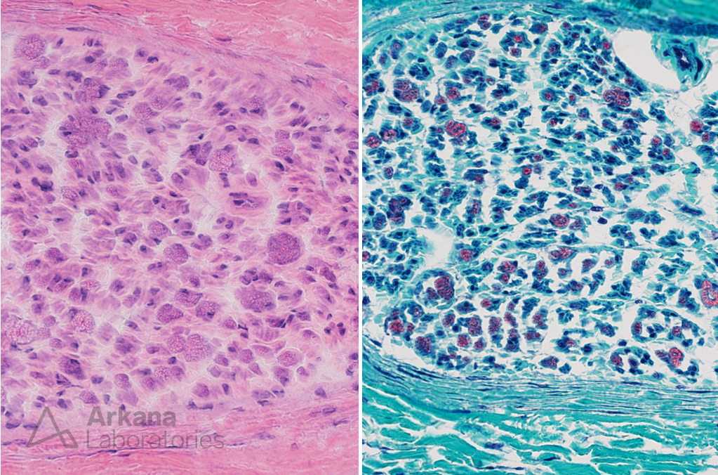
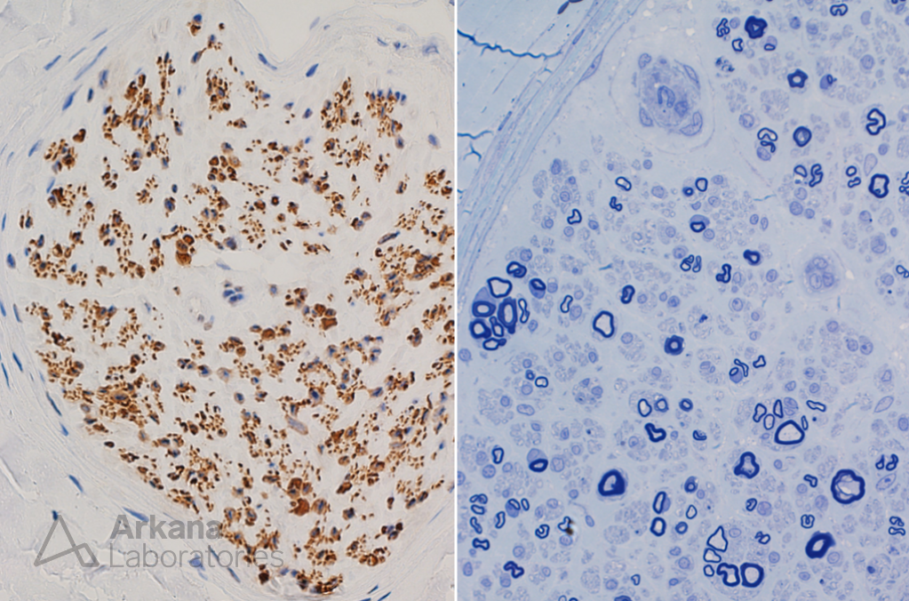
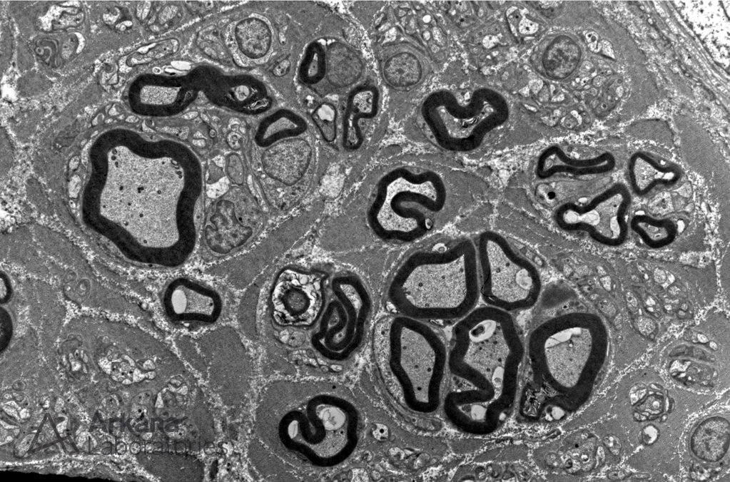
Figure 1: These high magnification images show a peripheral nerve fascicule in cross section. The myelinated axons are visible on H&E but are better demonstrated on modified Gomori Trichrome stain.
Figure 2: The high magnification image of the toluidine blue stained thick section (right) shows several clusters of myelinated axons. Note the variable thickness of the myelin sheath in the axons within the clusters. The clusters are also visible in the neurofilament immunohistochemically stained section (left). In retrospect, can you see the clusters on the H&E and Trichrome?
Figure 3: Electron microscopy nicely demonstrates an abnormal cluster of larger diameter myelinated axons.
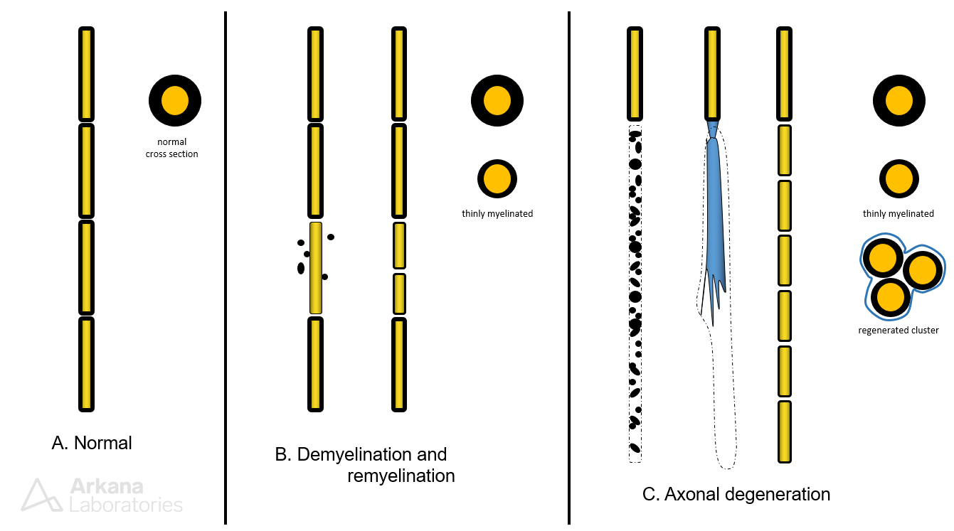
Correct answer: A. Regenerated clusters
- When a neuron and/or its axon is sub-lethally damaged the stump of the axon will attempt to reconnect to its target by sending out multiple “axonal sprouts.” These travel within the intact basement membrane sleeve that surrounds the associated myelinating Schwann cell (kind of like an axonal highway). These multiple sprouts may later become re-myelinated. It takes time to fully regain the thickness of an adult myelin sheath, and therefore remyelinated axons typically have thinner myelin sheaths. Please see explanatory image that follows.
- In this context, regenerated clusters are an indication of prior axonal injury and subsequent successful recovery.
Quick note: This post is to be used for informational purposes only and does not constitute medical or health advice. Each person should consult their own doctor with respect to matters referenced. Arkana Laboratories assumes no liability for actions taken in reliance upon the information contained herein.