Clinical history
This 20-year-old patient was found in a prolonged seizure state (status epilepticus of unknown duration). CPK levels at the time of hospital admission were greater than 200,000.
Figures #1 through #3 show which of the following?
A. Necrotic myofibers
B. Ragged red fibers
C. Normal muscle
D. Denervation

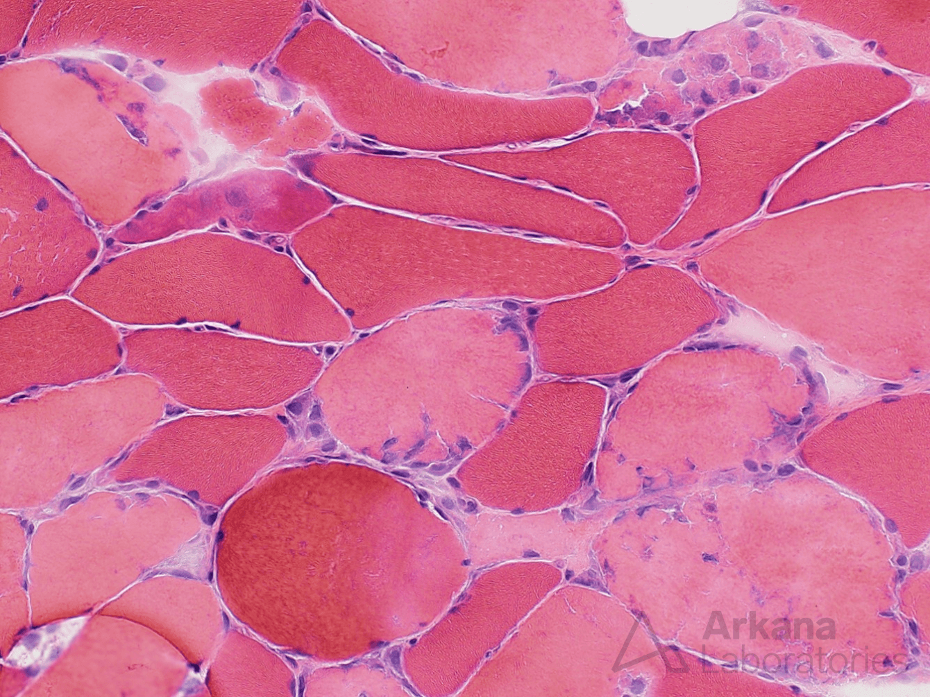

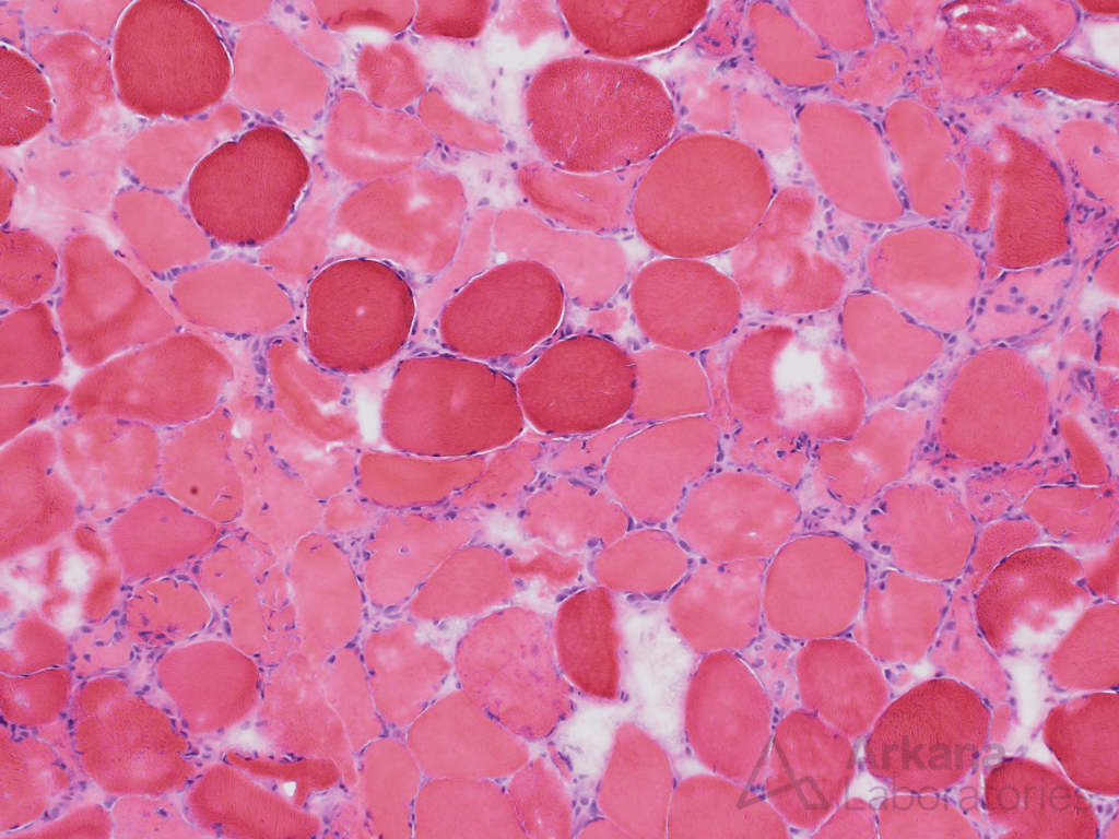
Figure 1: frozen section H&E 100x magnification
Medium magnification image showing numerous pale staining muscle fibers, without associated chronic inflammation.
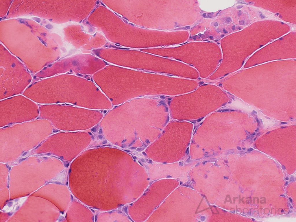
Figure 2: frozen section H&E 200x magnification
Higher magnification image showing pale staining muscle fibers with smudgy appearing nuclei and early macrophage infiltration.
Note that the pale staining muscle fibers all have a relatively similar appearance (“monophasic” pattern of myofiber injury).
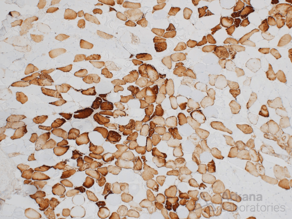
Figure 3: C5b-9 immunostain 40x magnification
Low magnification image showing numerous necrotic myofibers with sarcoplasmic immunostaining for C5b-9 (complement membrane attack complex).
Correct answer
A. Necrotic myofibers
The figures show numerous acutely necrotic muscle fibers. These have a monophasic appearance suggesting single recent episode of myofiber injury. In the context of this patient’s clinical history this is most likely related to prolonged muscle contraction due to seizure. Similar changes may be seen in toxic myopathy and viral myositis.
Quick note: This post is to be used for informational purposes only and does not constitute medical or health advice. Each person should consult their own doctor with respect to matters referenced. Arkana Laboratories assumes no liability for actions taken in reliance upon the information contained herein.