Clinical History
This 40-year-old female who presented with generalized muscle weakness, progressive ptosis and cognitive impairment.
Question
What of the following is the most likely diagnosis based on figures #1 and #2?
A. Polymyositis
B. IBM
C. Sarcoidosis
D. Mitochondrial myopathy
Answer – Mitochondrial Myopathy
The correct answer is D. Mitochondrial myopathy.
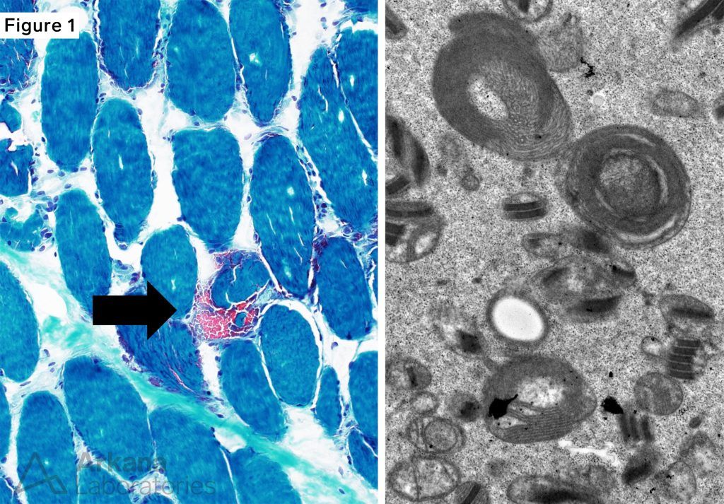
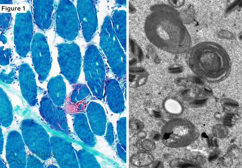
Figure 1: Modified Gomori Trichrome frozen section and EM
Modified Gomori Trichrome showed scattered muscle fibers with increased mitochondrial type staining, including occasional ragged-red muscle fibers (arrow).
Electron microscopy demonstrated frequent muscle fibers containing mitochondria with abnormal architecture (enlarged with spiral arrangement of crista) and/or containing paracrystalline inclusions.
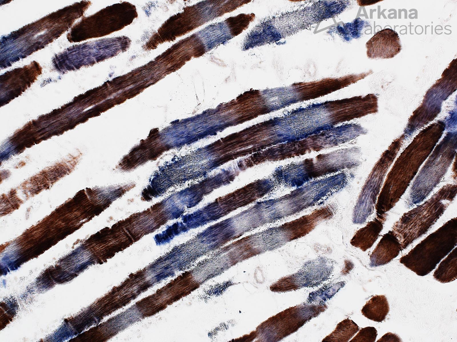
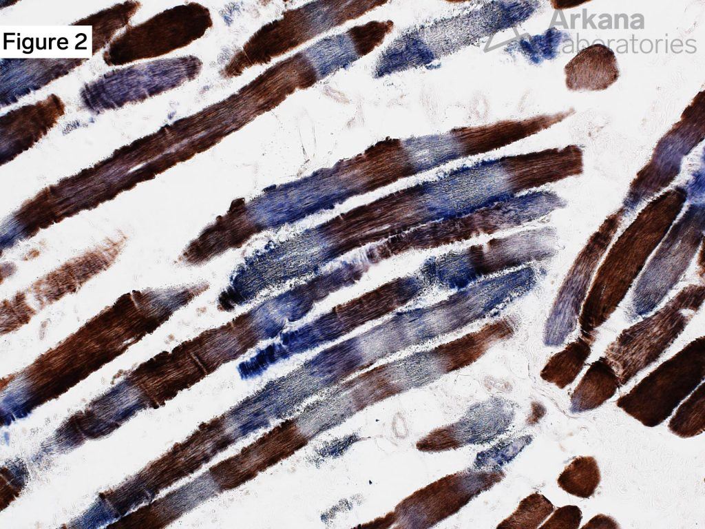
Figure 2: Frozen section combined COX-SDH
Combined COX-SDH enzyme histochemical stain showed frequent “COX-negative” muscle fibers (fibers which show absence of brown COX staining but intact blue SDH staining).
Note the striking segmental nature of the COX-negative staining in this case.
Mitochondrial Myopathy
- The cells of your body coordinate two genomes: the nuclear genome (nDNA) which encodes SDH and the mitochondrial genome (mtDNA) which encodes COX.
- Recent research has shown that there are different populations of mitochondria in muscle (subsarcolemmal and intermyofibrillar) and that these form and interconnecting network. The segmental pattern of COX-negative staining is thought to possibly reflect a biologic safety mechanism of the mitochondrial “power grid.” Please see reference for additional information.
Reference(s)/Additional Reading:
-
Willingham TB, Ajayi PT, Glancy B. Subcellular Specialization of Mitochondrial Form and Function in Skeletal Muscle Cells. Front Cell Dev Biol. 2021 Oct 15;9:757305. doi: 10.3389/fcell.2021.757305. PMID: 34722542; PMCID: PMC8554132.
- Picard M, White K, Turnbull DM. Mitochondrial morphology, topology, and membrane interactions in skeletal muscle: a quantitative three-dimensional electron microscopy study. J Appl Physiol (1985). 2013 Jan 15;114(2):161-71. doi: 10.1152/japplphysiol.01096.2012. Epub 2012 Oct 25. PMID: 23104694; PMCID: PMC3544498.
- Glancy B, Hartnell LM, Combs CA, Femnou A, Sun J, Murphy E, Subramaniam S, Balaban RS. Power Grid Protection of the Muscle Mitochondrial Reticulum. Cell Rep. 2017 Apr 18;19(3):487-496. doi: 10.1016/j.celrep.2017.03.063. Erratum in: Cell Rep. 2018 May 29;23(9):2832. PMID: 28423313; PMCID: PMC5490369.
Quick note: This post is to be used for informational purposes only and does not constitute medical or health advice. Each person should consult their own doctor with respect to matters referenced. Arkana Laboratories assumes no liability for actions taken in reliance upon the information contained herein.