This 60 year-old patient presented with distal asymmetric muscle weakness and normal CPK.
Which of the following abnormalities is well-demonstrated in this patient’s muscle biopsy with this stain?
A. Fiber type grouping
B. Tubular aggregates
C. Rimmed vacuoles
D. Ring fibers
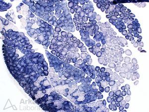
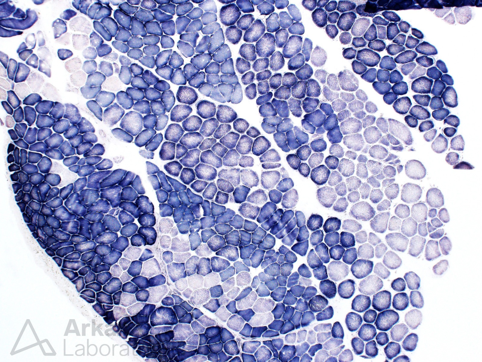
Figure 1: Succinic acid dehydrogenase (SDH) enzyme histochemical stain 100x original magnification
Answer: Fiber type grouping
Succinic acid dehydrogenase (SDH) enzyme histochemical stain highlights mitochondria. As a result, type 1 muscle fibers (oxidative/slow twitch) which contain more mitochondria stain more darkly than type 2 fibers (glycolytic/fast twitch). This patient’s muscle biopsy shows fiber grouping (loss of normal “checkerboard” distribution of type 1 and 2 myofibers), a feature indicating prior denervation and subsequent successful reinnervation.
While fiber typing in muscle biopsy samples is best performed with ATPase enzyme histochemical staining (pHs 9.6, 4.6, and 4.3) or immunohistochemical staining for myosin heavy chain (fast or slow isoforms), differences in type 1 and type 2 fiber type staining can also be appreciated on other stain preparations routinely used in evaluation of muscle biopsies.
Quick note: This post is to be used for informational purposes only and does not constitute medical or health advice. Each person should consult their own doctor with respect to matters referenced. Arkana Laboratories assumes no liability for actions taken in reliance upon the information contained herein.