This 75-year-old patient presented with complaints of numbness, tingling and burning sensation involving his right lower extremity below the level of the knee (stocking-like distribution) and foot drop. The patient’s past medical history is significant for coronary artery disease, impaired fasting glucose, peripheral neuropathy, and hypertension.
In addition to diffuse thickening of the perineurium seen in Figure #1, what abnormality is seen in Figures #2 – #4?
A. Microfascicles
B. Angioma
C. Schwannoma
D. Neurofibroma
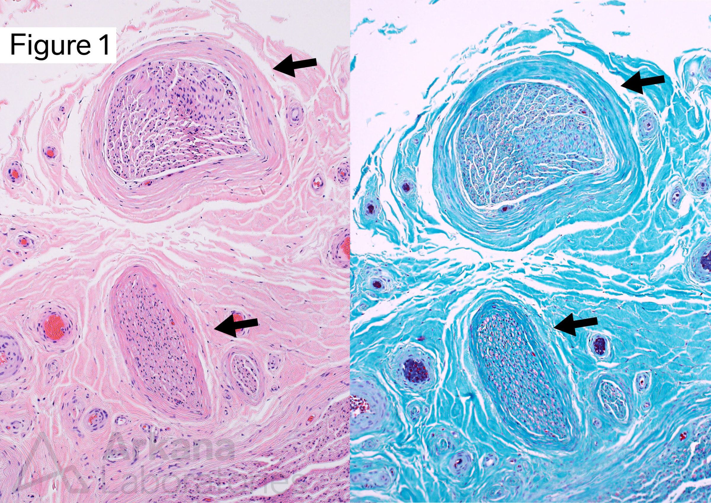
Two peripheral nerve fascicles show diffuse moderate to marked thickening of the perineurium (arrows).
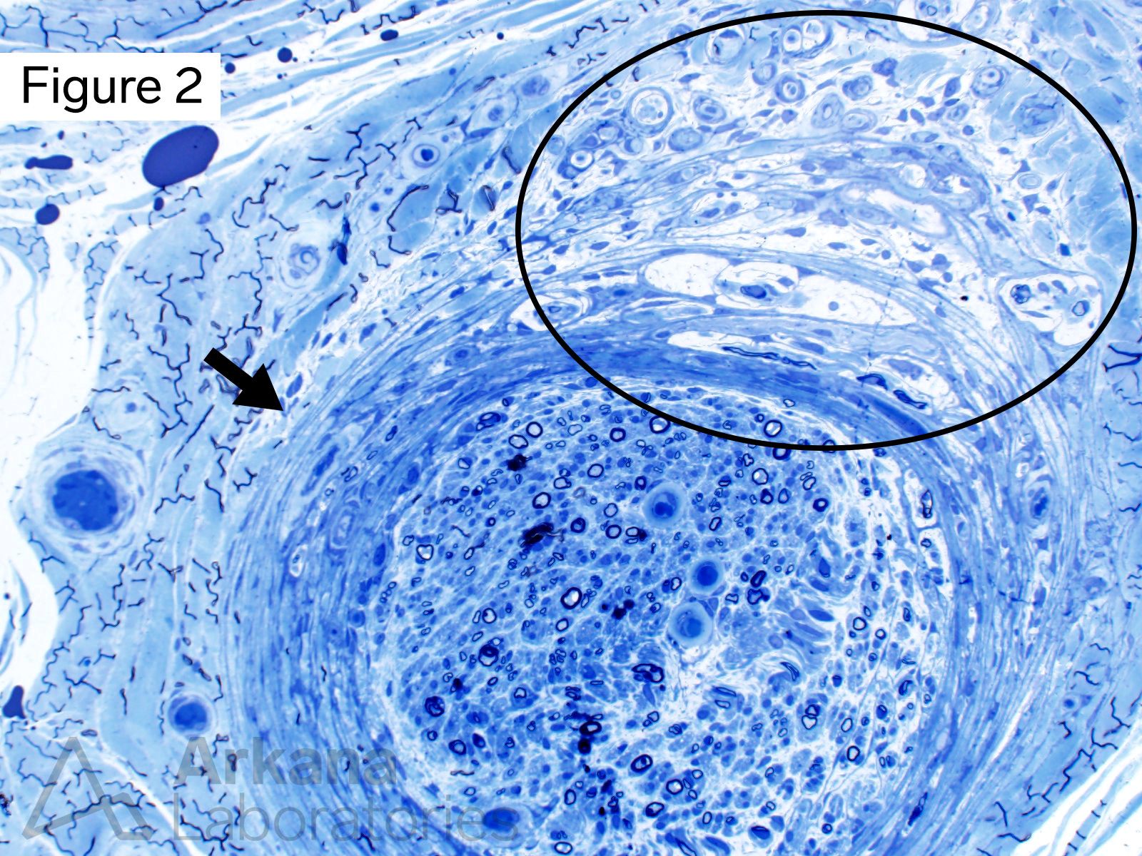
In addition to diffuse thickening of the perineurium (arrow) this nerve fascicle shows microfascicles within the perineurium and adjacent epineurium (circled area). These are seen at higher magnification in figures #3 and #4. Note: the dark squiggles seen toward the left upper and lower side of this image represent fine wrinkles in the tissue section (i.e. artifact).
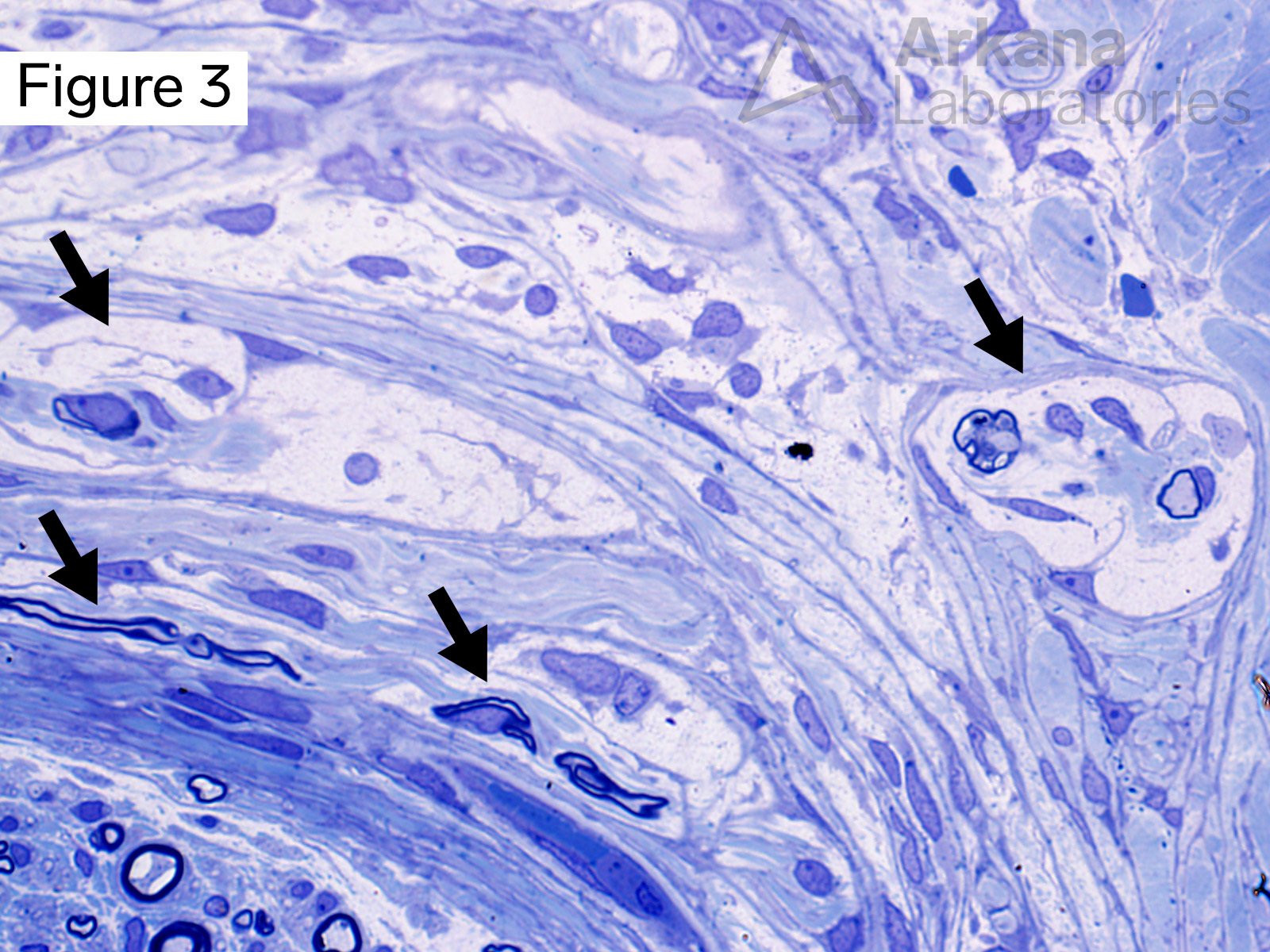
Higher magnification showing microfascicles (arrows) within the perineurium and epineurium. Some of the microfascicles contain myelinated axons. The myelinated axons shown in the lower left corner of this image are in their normal location within the nerve fascicle.
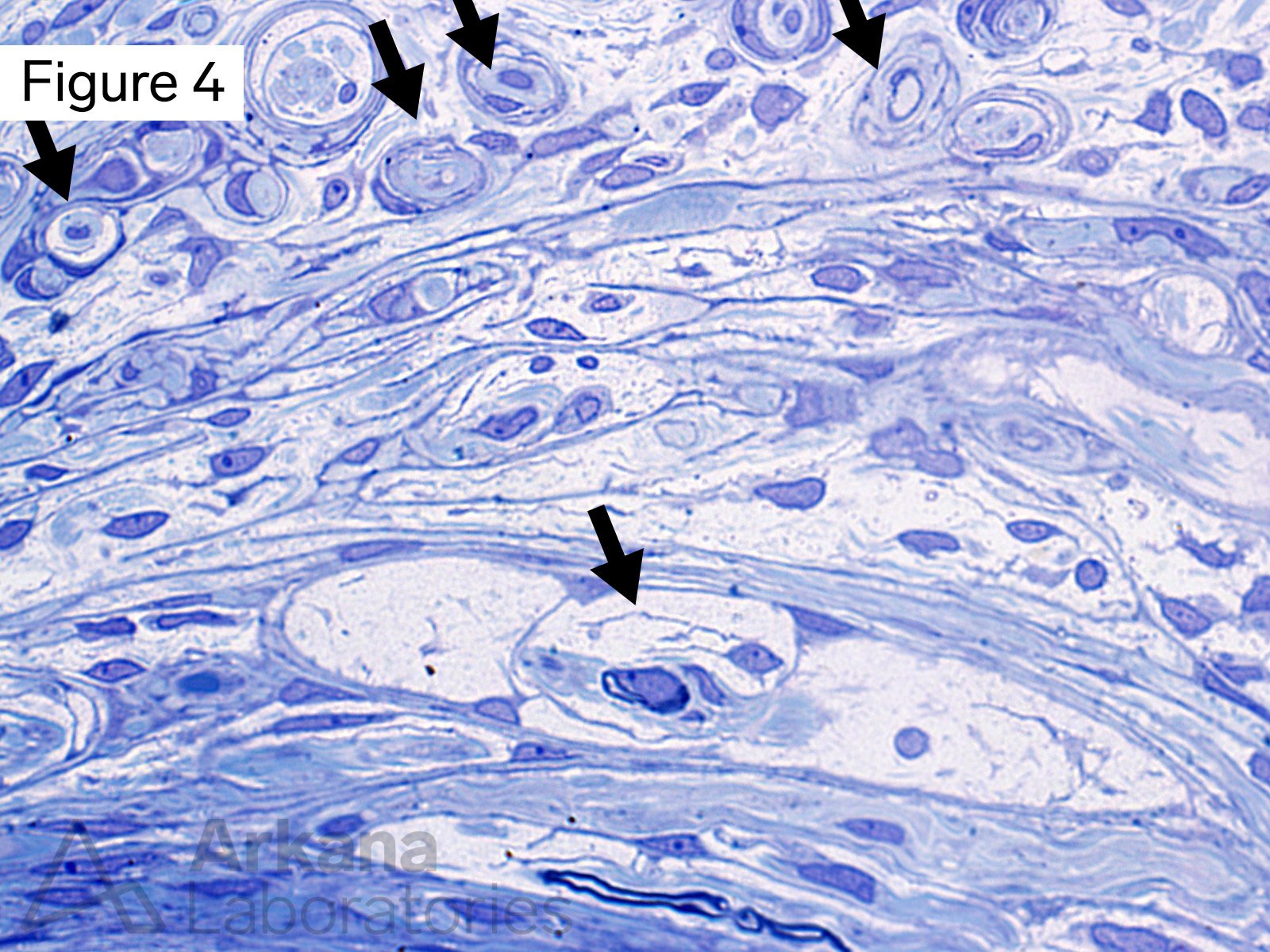
Higher magnification showing microfascicles within the perineurium and epineurium (arrows). Many of these do not show obvious myelination.
Answer: Microfascicles
The toluidine blue stained thick sections show the focal presence of microfascicles. Microfascicles are comprised of unmyelinated or myelinated axons surrounded by a layer(s) of fibroblasts and perineurial cells, and represent a disorganized healing response. They are seen in the setting of peripheral nerve injury (disruption of the perineurium) following surgery (traumatic or amputation neuroma), but may also be seen in the context of nerve injury related to vasculitis, diabetic vasculopathy, and perineuritis (for example leprosy).
References/Additional Reading
Zabaglo M, Dreyer MA. Neuroma. [Updated 2022 Jan 9]. In: StatPearls [Internet]. Treasure Island (FL): StatPearls Publishing; 2022 Jan-. Available from: https://www.ncbi.nlm.nih.gov/books/NBK549838/
Oliveira KMC, Pindur L, Han Z, Bhavsar MB, Barker JH, Leppik L. Time course of traumatic neuroma development. PLoS One. 2018 Jul 16;13(7):e0200548. doi: 10.1371/journal.pone.0200548. PMID: 30011306; PMCID: PMC6047790.
Dyck PJ, Norell JE, Dyck PJ. Microvasculitis and ischemia in diabetic lumbosacral radiculoplexus neuropathy. Neurology. 1999 Dec 10;53(9):2113-21. doi: 10.1212/wnl.53.9.2113. PMID: 10599791.
Antunes SL, Medeiros MF, Corte-Real S, Jardim MR, Nery JA, Hacker MA, Valentim Vda C, Amadeu TP, Sarno EN. Microfasciculation: a morphological pattern in leprosy nerve damage. Histopathology. 2011 Jan;58(2):304-11. doi: 10.1111/j.1365-2559.2011.03749.x. PMID: 21323955.
Quick note: This post is to be used for informational purposes only and does not constitute medical or health advice. Each person should consult their own doctor with respect to matters referenced. Arkana Laboratories assumes no liability for actions taken in reliance upon the information contained herein.