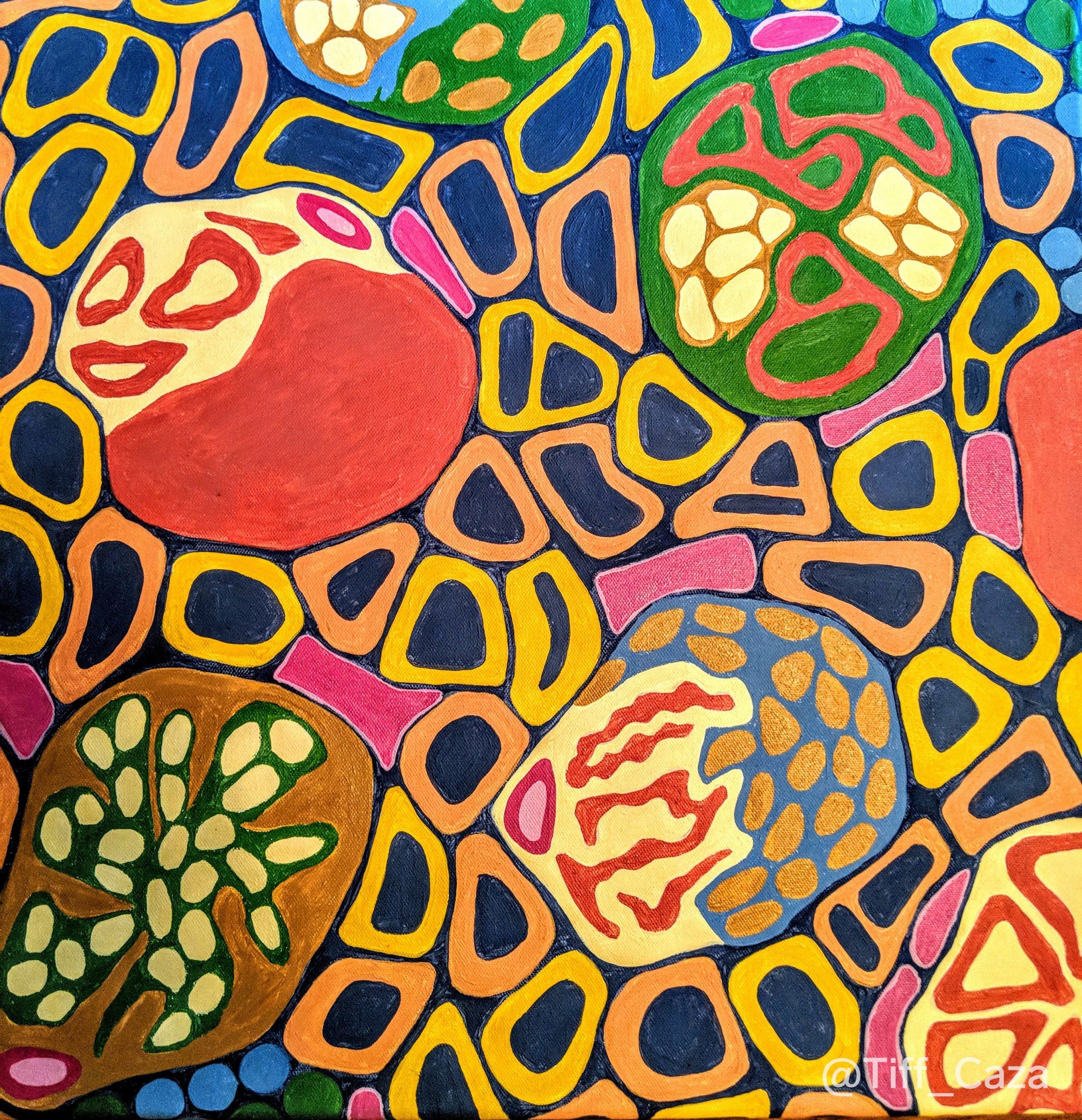
The above painting shows glomeruli with mesangial hypercellularity, endocapillary hypercellularity, and crescent formation. These findings can be seen in IgA nephropathy, and other active glomerulonephritides. These lesions shown in the painting above are represented in the Oxford Classification for IgA nephropathy. The Oxford classification is a scoring system on kidney biopsies that includes mesangial hypercellularity (M0 = <50%, M1 = >50%), endocapillary hypercellularity (E0 = none, E1 = present), segmental sclerosis (S0 = absent, S1 = present), tubular atrophy / interstitial fibrosis (T0 = <25%, T1 25-50%, T2 >50%), and crescents (C0 = absent, C1 = up to 25%, C2 = greater than 25%). The Oxford classification has been validated internationally and is used within current prognostication tools to predict outcomes at up to 5 years post-diagnosis (Barbour et al, 2019).
IgA Nephropathy
IgA nephropathy is diagnosed by the presence of at least 2 + granular mesangial IgA deposits within glomeruli by immunofluorescence, with or without the proliferative and chronic changes seen by light microscopy, as described above. Primary IgA nephropathy is thought to be due to antibodies against galactose deficient IgA1 depositing within glomeruli. Secondary causes of IgA nephropathy include cirrhosis and hepatobiliary disorders, psoriasis, inflammatory bowel disease, celiac disease, and infections (especially Staphylococcal infections, making up an IgA dominant infection-associated glomerulonephritis).
Differentiating primary IgA nephropathy from IgA dominant infection-associated glomerulonephritis is critical. There are treatment implications (treatment of the underlying infection rather than immunosuppression), and prognostic implications (IgA dominant infection-associated glomerulonephritis often has a poor prognosis with progression to end stage renal disease in a majority of patients). Since primary IgA nephropathy is believed to be caused by underglycosylation of O-glycans in the hinge region of IgA1, detection of underglycosylated IgA was investigated to separate out patients with primary IgA nephropathy versus secondary causes of IgA deposition within glomeruli.
Two recent case series (each with 75-100 patients) have shown positive anti-galactose deficient (Gd)-IgA staining using the KM55 monoclonal antibody on renal biopsies with primary IgA nephropathy. However, there are issues with sensitivity (Charu et al, 2019) and specificity (Cassol et al, 2019) using Gd-IgA as a marker. In the recent case series from Cassol et al, 69 percent of patients with IgA deposition within glomeruli due to infections had some positive Gd-IgA staining. In the series from Charu et al, a cutoff of 50% of positive glomeruli was used to improve specificity.
However, KM55 staining was insensitive, with a subset of negative cases. While there may be some value in use of this antibody as an adjunctive tool, it cannot definitively separate out primary versus secondary forms of IgA nephropathy in all cases. Currently, the KDIGO guidelines recommend assessing all patients with biopsy-proven IgA nephropathy for secondary causes, to exclude infections, liver disease, and gastrointestinal disease as potential etiologies.
References:
Barbour SJ, Coppo R, Zhang H, Liu Z-H, Suzuki Y, Matsuzaki K, Karafuchi R, Er L, Espino-Hernandez G, Kim SJ, Reich HN, Feehally J, Cattran DC, and the International IgA Nephropathy Network. Evaluating a New International Risk-Prediction Tool in IgA Nephropathy. JAMA Internal Medicine 2019; published ahead of print online April 13, 2019.
Cassol CA, Bott C, Nadasdy GM, Alberton V, Malvar A, Nagaraja HN, Nadasdy T, Rovin BH, Satoskar AA. Immunostaining for galactose deficient IgA is not specific for primary immunoglobulin A nephropathy. Nephrology Dialysis Transplant 2019; Aug 1 e-pub ahead of print.
Charu V, Best-Rocha A, Sharma S, Larsen C. KM55 is a highly specific but insensitive marker for IgA nephropathy. Abstracts from the United States and Canadian Academy of Pathology 2019 scientific meeting: medical renal pathology (1581-1620; abstract #1591); Laboratory Investigation 2019 Mar; 99 (1): 1-36.
Quick note: This post is to be used for informational purposes only and does not constitute medical or health advice. Each person should consult their own doctor with respect to matters referenced. Arkana Laboratories assumes no liability for actions taken in reliance upon the information contained herein.

