Small destructive B cell or plasma cell clones may be responsible for disease development.
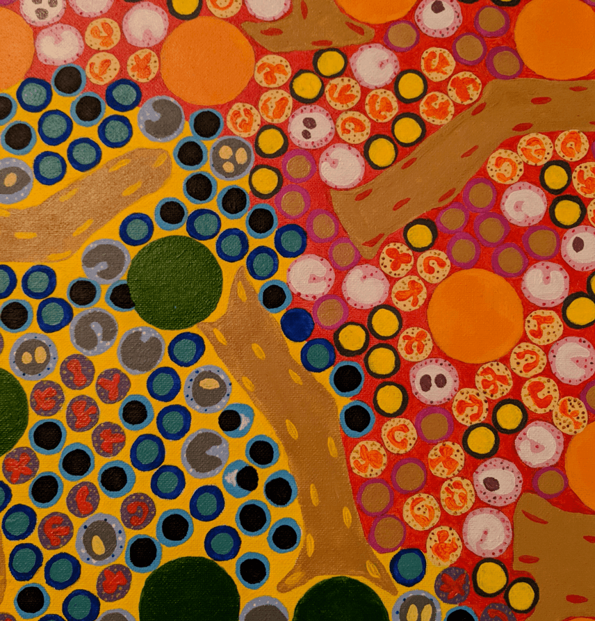
The above painting shows normal bone marrow, containing erythroid and myeloid precursors at various stages of maturation and normal trabeculae. Bone marrow biopsies may be performed to evaluate for a lymphoid or plasma cell clone in patients with monoclonal gammopathies of undetermined significance (MGUS) and in work-up of patients found to have a monoclonal gammopathy of renal significance (MRGS).
Monoclonal gammopathies of renal significance occur when light chain restriction within a kidney biopsy is thought to be responsible for the patient’s renal dysfunction, which is usually acute kidney injury or proteinuria. This includes a broad spectrum of findings, which includes but is not limited to, light chain deposition disease, light chain proximal tubulopathies (with and without crystals), amyloidosis, and proliferative glomerulonephritis with monoclonal immunoglobulin deposits (PGMID). Light chain cast nephropathy is considered a myeloma-defining lesion and is not under the spectrum of a monoclonal gammopathy of renal significance. In patients with PGMID, bone marrow biopsies may be normal and not reveal a pathogenic plasma cell clone, despite being driven by a clonal process. Small destructive clones, below the limits of detection, are thought to be responsible in such cases.
In cases of PGMID, there is a low overall detection rate of circulating monoclonal paraproteins (approximately 37%), as determined by serum protein electrophoresis, serum immunofixation, and serum free kappa-to-lambda light chain ratios, as well as urine protein electrophoresis and urine immunofixation (Gumber et al, 2018). Bone marrow biopsy of patients with PGMID have a similar rate of detection of a pathogenic plasma cell or lymphoid clone (32%), through use of flow cytometry capturing numerous cellular events (such as used for evaluation of minimal residual disease).
Cases with a membranoproliferative pattern of glomerulonephritis with monoclonal immunoglobulin deposits have a slightly increased rate of detection of a pathogenic plasma cell clone. While uncommon, the light chain only variant of PGMID has a much higher rate of detection of a pathogenic clone. There is 65-73% detection on serum and urine immunofixation, 83% by serum free light chain assay, and 88% on bone marrow biopsy, as identified in a recent series of 17 patients (Nasr et al, 2020).
Despite the absence of detection of a pathogenic clone, there is a good response to clone directed therapies in patients with PGMID. Empiric treatment of patients with a presumed underlying clone led to an overall renal response rate of 88% (Gumber et al, 2018), with a subset of patients achieving complete remission (38% with <500 mg proteinuria per 24 h collection). All patients treated with clone based approaches did not progress to end stage kidney disease at an average of 2 years of follow up, although long-term outcomes are not yet known.
While a hematologic workup is necessary in patients with PGMID, use of clone-directed approaches may prove helpful, whether or not a clone is identified. Identification of an underlying clone can help determine an appropriate therapy, and know whether to start with a B-cell or plasma cell-directed approach.
References:
- Gumber R, Cohen JB, Palmer MB, Kobrin SM, Vogl DT, Wasserstein AG, Nasta SD, Bleicher MB, Bloom RD, Dember L, Cohen A, Weiss BM, Hogan JJ. A clone-directed approach may improve diagnosis and treatment of proliferative glomerulonephritis with monoclonal immunoglobulin deposits. Kidney International 2018 July; 94 (1): 199-205.
- Nasr SH, Larsen CP, Sirac C, Theis JD, Domenger C, Chauvet S, Javaugue V, Hogan JJ, Said SM, Dasari S, Vrana JA, McPhail ED, Cornell LD, Vilaine E, Massy ZA, Boffa J-J, Buob D, Toussaint S, Guincestre T, Touchard G, D’ Agati VD, Leung N, Bridoux F. Light chain only variant of proliferative glomerulonephritis with monoclonal immunoglobulin deposits is associated with a high detection rate of the pathogenic plasma cell clone. Kidney International 2020; in press.
Quick note: This post is to be used for informational purposes only and does not constitute medical or health advice. Each person should consult their own doctor with respect to matters referenced. Arkana Laboratories assumes no liability for actions taken in reliance upon the information contained herein.
