
Clinical presentation
A 65-year-old male presented with new onset anasarca and nephrotic syndrome. The patient has a history of hypertension but was otherwise healthy with no significant past medical history. Further evaluation revealed the presence of anemia, with a hemoglobin level of 8.5 g/dL. The patient also had an elevated serum creatinine of 1.8 mg/dL and a serum albumin of 1.6 g/dL. Urinalysis showed the presence of proteinuria and blood in the urine. A 24-hour urine collection showed a protein-to-creatinine ratio of 10 g/g. A biopsy of the patient’s kidney was performed.
Light Microscopy
The key observation revealed by light microscopy is depicted in the images below (PAS and Jones silver stains).
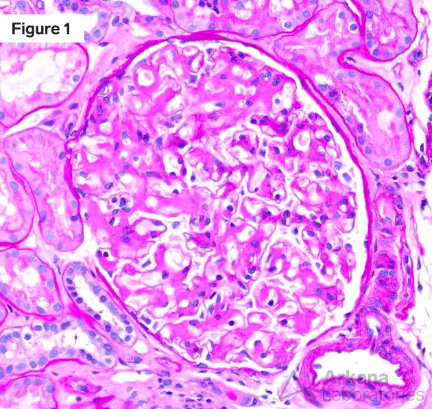
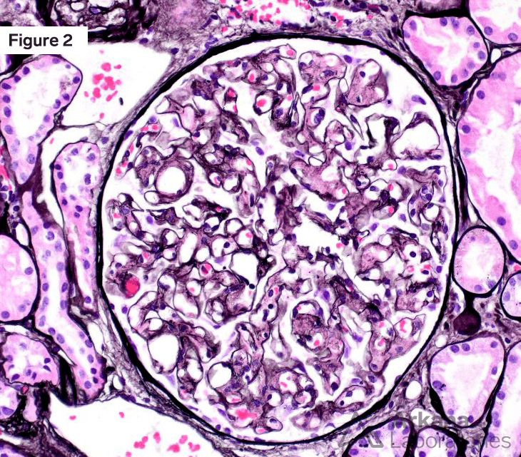
Microscopic examination of the glomeruli revealed an increased mesangial area with a weakly PAS-positive, non-argyrophilic amorphous material that stains positive for Congo red. When viewed under polarized light, the material exhibits apple-green birefringence.
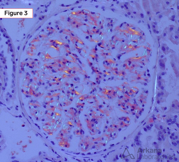
Congo red
Additional Clues
The figure below shows the immunofluorescence findings. The amorphous material seen in the image stains positively for lambda light chains (on the right) and negatively for kappa light chains (on the left). It’s worth noting that similar amorphous material, which is also lambda positive, can be found in the interstitium and small vessels, though these areas are not shown in the figure.
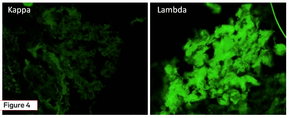
IF
Ultrastructural examination of a glomerulus by electron microscopy showed the deposition of small randomly arranged fibrils within the mesangium and extending into capillary walls.
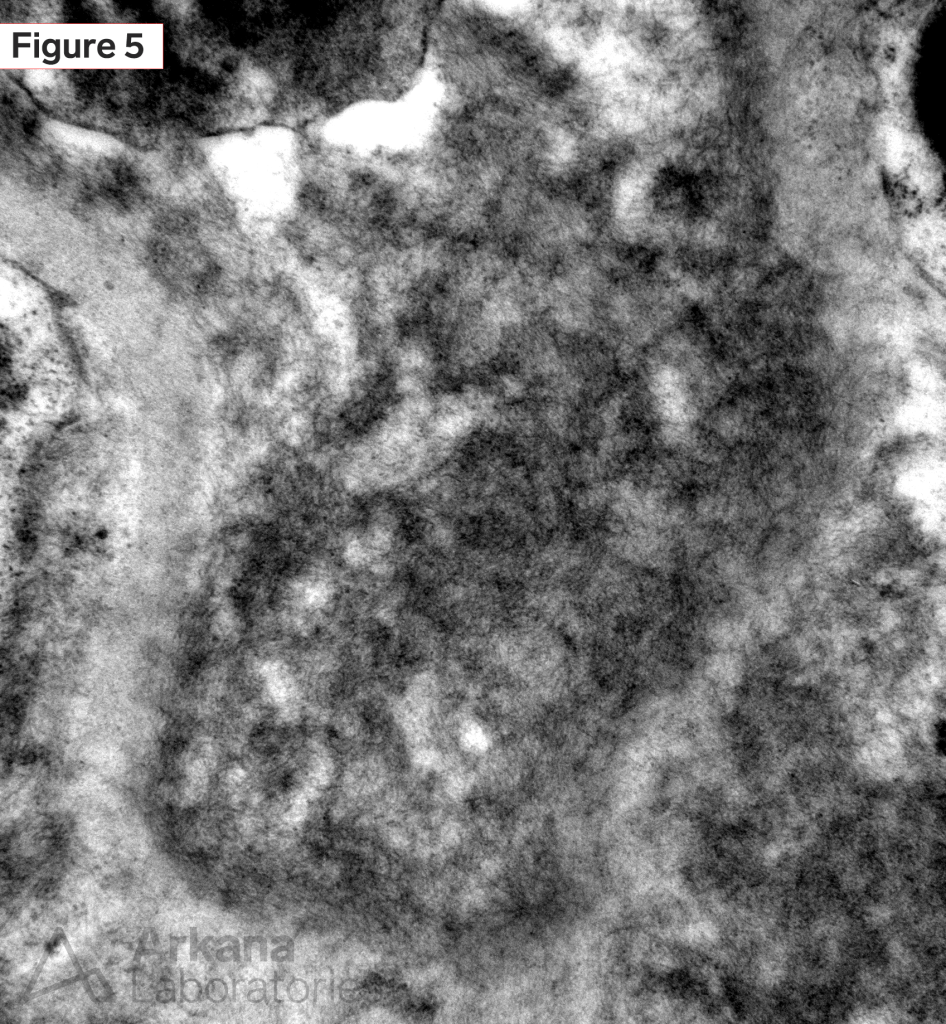
EM
Diagnosis
The kidney biopsy revealed the presence of amyloid deposits, derived from lambda-type immunoglobulin light chains. This confirmed a diagnosis of AL amyloidosis, the most common type of renal amyloidosis in the USA. AL amyloidosis occurs when abnormal light chains produced by plasma cells accumulate in tissues and form amyloid deposits. In this case, the patient had lambda light chain deposition, which is present in about 75% of AL amyloidosis. The amyloid deposits can affect all renal compartments, with glomerular involvement being common, leading to proteinuria. Approximately 20% of AL amyloidosis is associated with multiple myeloma, leukemia, or lymphoma. Cardiac involvement in AL amyloidosis can also result in congestive heart failure, which is a major factor affecting survival.
References
Colvin, Robert, B. and Anthony Chang. Diagnostic Pathology: Kidney Diseases. Available from: Elsevier eBooks+, (3rd Edition). Elsevier – OHCE, 2019.
Jennette JC, Olson JL, Silva FG, D’Agati V, Heptinstall RH *1920 *, eds. Heptinstall’s Pathology of the Kidney: Includes Interactive EBook with Complete Content. Seventh edition. Wolters Kluwer; 2015.
Quick note: This post is to be used for informational purposes only and does not constitute medical or health advice. Each person should consult their own doctor with respect to matters referenced. Arkana Laboratories assumes no liability for actions taken in reliance upon the information contained herein.