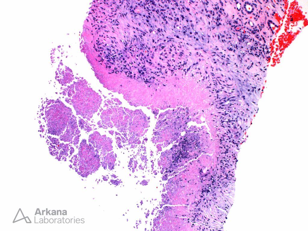
Incidental findings on renal biopsies are not uncommon. The presented image shows a caseating granuloma within the renal medulla of a diabetic patient without known history of infection. The lesion shows central necrosis surrounded by palisading histiocytes and scattered lymphocytes. Caseating granuloma are highly suspicious for mycobacterial or fungal infections. While AFB, Auramine-Rhodamine and GMS stains are negative in this case, due to the relative insensitivity of these stains, ruling out an infection clinically is warranted. In those cases with non-caseating granulomata, the differential diagnosis would expand to include drug reactions, sarcoidosis and other autoimmune disorders. In such cases, performing histochemical stains for microorganisms would also be of value to help rule out infectious etiologies.
Quick note: This post is to be used for informational purposes only and does not constitute medical or health advice. Each person should consult their own doctor with respect to matters referenced. Arkana Laboratories assumes no liability for actions taken in reliance upon the information contained herein.