This is the H&E from the frozen tissue submitted for IF examined under fluorescent light. In a patient with anemia, thrombocytopenia, high LDH and schistocytes, what is your diagnosis?
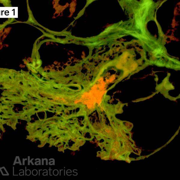
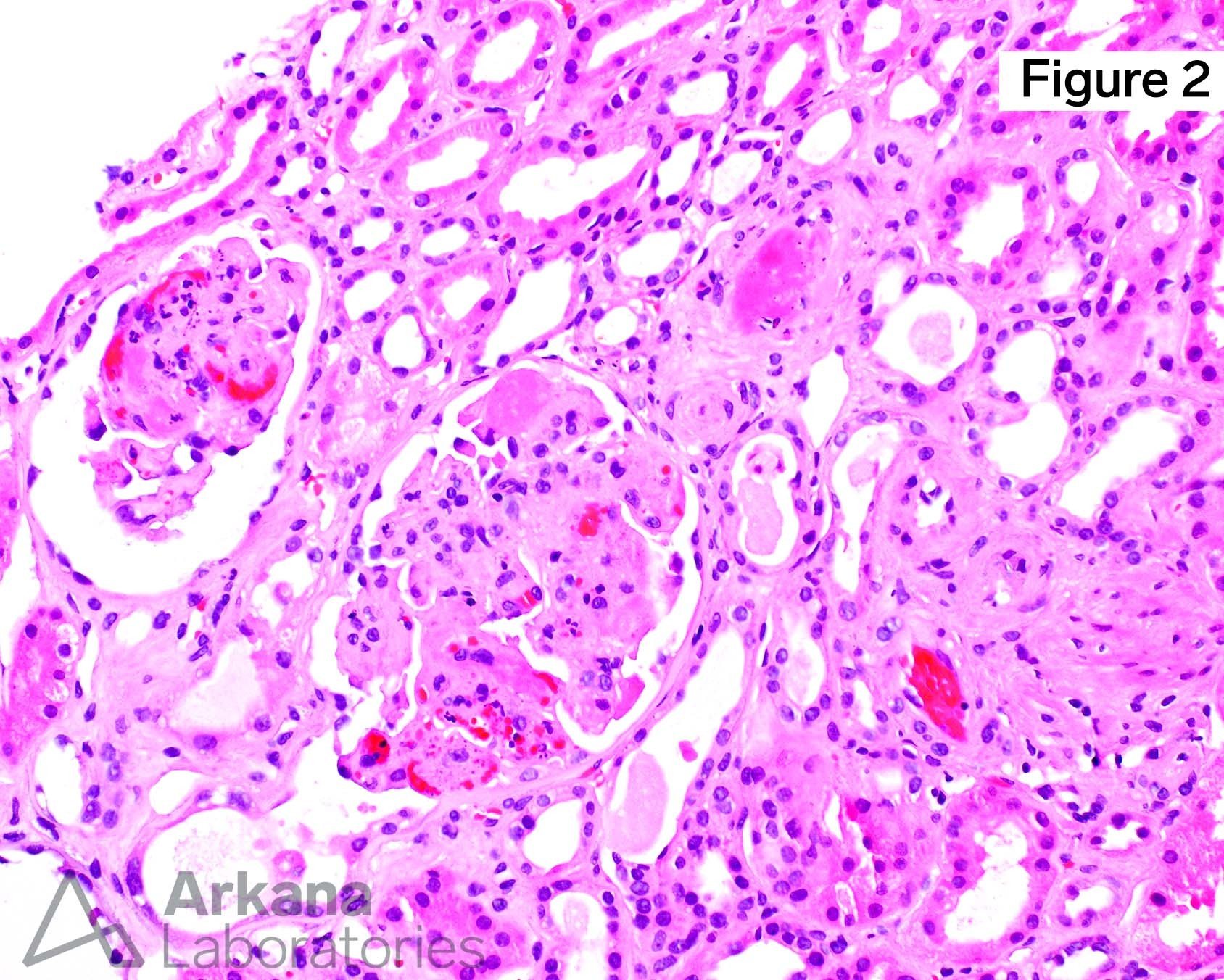
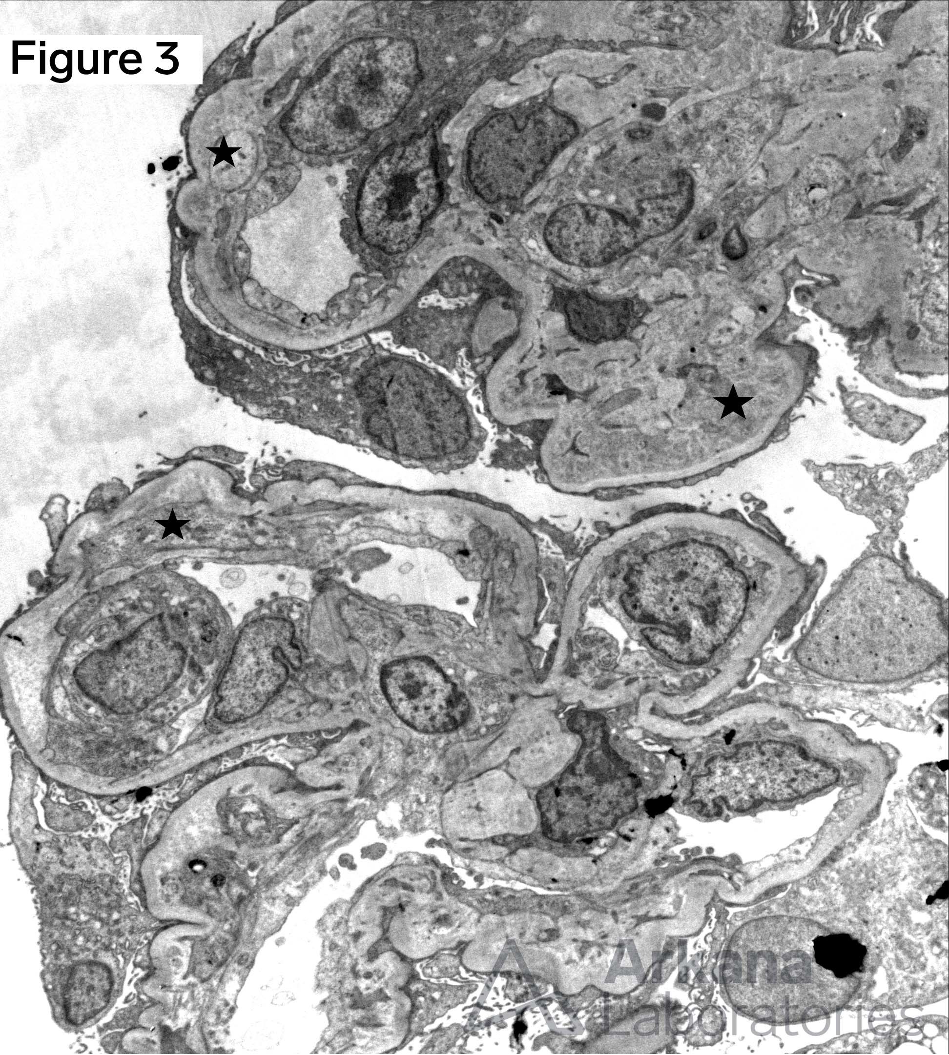
The image shows a fibrin thrombus located at the hilum of the glomerulus. This finding is consistent with thrombotic microangiopathy. Light microscopy demonstrated numerous glomerular and arteriolar fibrin thrombi (Figure 2). By electron microscopy, subendothelial lucent widening and new glomerular basement membrane formation were present (stars, Figure 3).
Quick note: This post is to be used for informational purposes only and does not constitute medical or health advice. Each person should consult their own doctor with respect to matters referenced. Arkana Laboratories assumes no liability for actions taken in reliance upon the information contained herein.