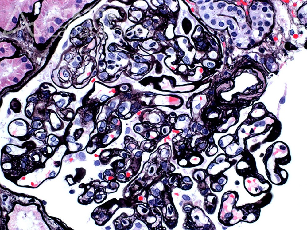
What are the two major changes seen in this image from an allograft kidney and what are these findings concerning for?
Answer: The light microscopic image of a glomerulus on silver stain demonstrates glomerulitis as characterized by capillary loop occlusion by mononuclear cells and neutrophils along with endothelial enlargement which is concerning for active antibody-mediated rejection. Other features used to diagnose antibody-mediated rejection, in this case, included severe peritubular capillaritis and diffusely positive C4d in peritubular capillaries. The image also shows chronic changes of antibody-mediated rejection with multiple loops showing double contour formation. For more information on the Banff criteria for transplant rejection, see the following reference:
Reference: Loupy A, Haas M, Roufosse C, et al. The Banff 2019 Kidney Meeting Report (I): Updates on and clarification of criteria for T cell– and antibody-mediated rejection. Am J Transplant. 2020; 00: 1–14.
Quick note: This post is to be used for informational purposes only and does not constitute medical or health advice. Each person should consult their own doctor with respect to matters referenced. Arkana Laboratories assumes no liability for actions taken in reliance upon the information contained herein.

