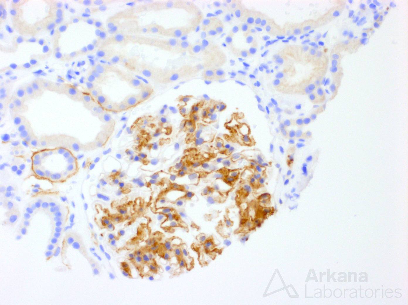
The patient is a 67 y/o Caucasian female with slowly progressive kidney disease and 2.3 gm/24 hr proteinuria. Aside from hypertension, she has no significant past medical history. Congo red was negative, however, immunofluorescence showed IgG and lambda staining within glomeruli. What is this immunoperoxidase stain?
For bonus points, what was found on electron microscopy?
The clinical vignette and findings raise a differential diagnosis of paraprotein-related glomerulopathy, fibrillary glomerulopathy, and type I cryoglobulinemia among others. The immunoperoxidase stain shown is a DNAJB9 stain that is specific and sensitive for fibrillary glomerulopathy. And, by electron microscopy, we would expect to find non-branching, randomly oriented fibrils within the mesangium and focally extended into the capillary walls. The foil in this question is the lambda light chain restriction. However, this finding is encountered in fibrillary glomerulopathy in approximately 10% of cases. And, while it may necessitate a paraproteinemia workup, in those with no paraproteinemia it is likely clinically insignificant.
Quick note: This post is to be used for informational purposes only and does not constitute medical or health advice. Each person should consult their own doctor with respect to matters referenced. Arkana Laboratories assumes no liability for actions taken in reliance upon the information contained herein.