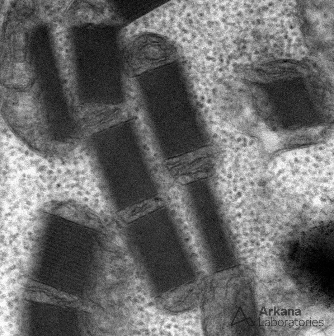
The image shows numerous crystalline-type mitochondrial inclusions. Morphologic abnormalities of mitochondria include abnormal size (usually enlargement), branched or concentric cristae, and various types of inclusions. The crystalline inclusions shown here usually take the form of rectangular arrays that occupy the entire width or length of a mitochondrion, and they may show a connection to the inner mitochondrial membrane. Such inclusions, like the other abnormalities listed above, are not specific for a particular mitochondrial disease, but they represent a structural clue to the possibility of an underlying metabolic disorder. Manifestations of kidney disease in patients with mitochondrial disease most commonly include tubular defects (e.g. proximal tubulopathy with Fanconi syndrome), but a growing number of glomerular diseases, often associated with FSGS lesions, have been described (e.g. coenzyme Q10 deficiency).
Reference:
Emma F, Montini G, Parikh SM, Salviati L. Mitochondrial dysfunction in inherited renal disease and acute kidney injury. Nat Rev Nephrol. 2016 May;12(5):267-80.
Quick note: This post is to be used for informational purposes only and does not constitute medical or health advice. Each person should consult their own doctor with respect to matters referenced. Arkana Laboratories assumes no liability for actions taken in reliance upon the information contained herein.