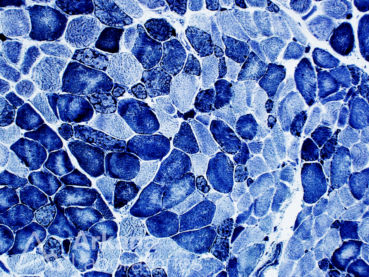
Figure 1: Hereditary myopathy with lobulated muscle fibers in an elderly male presenting with myalgias. The photomicrograph shows snap frozen skeletal muscle stained with the NADH-TR preparation demonstrating variably sized variably well-developed muscle fibers with irregular basement membrane contours and myofibrillar disorganization due to small triangular or wedge-shaped subsarcoplasmic areas of intensely dark staining character, consistent with small accumulations of mitochondria (also known as so-called “lobulated muscle fibers”). Some of the lobulated (trabeculated) muscle fibers are in small groups or scattered as single individual myofibers throughout the specimen. Original magnification: A. NADH-TR, 100.
Considering the clinical vignette from last week: An elderly male patient presents with chief complaint of “dull achy pain” predominantly in his thighs and hip girdle region. Muscle enzymes showed CPK 934. Other past medical history includes diabetes mellitus, hyperlipidemia, hypertension, hypercalcemia, and vitamin D deficiency. Due to clinical concern for a possible acquired inflammatory myopathy, such as polymyositis, the decision was made to biopsy the left thigh. The muscle biopsy shows lobulated muscle fibers, see Figure 1.
What are a few of the hereditary etiologies for myopathy with lobulated muscle fibers?

Answer: All of the Above
If you chose answer choice (D), all of the above, then you’re correct! Lobulated (trabeculated) muscle fibers are not entirely specific as to etiology, and may be seen in hereditary / genetic and acquired conditions (see prior week’s #NeuroNotes). See references below.
Lobulated muscle fibers are not entirely specific to one clear cut defined myopathic process; however, there is an associated with calpainopathy – that is to say limb-girdle muscular dystrophy associated with calpain-3 deficiency (LGMD 2A), which is an autosomal recessive disease.
These muscle fibers have also been described in other myopathic processes, such as facioscapulohumeral dystrophy (FSHD), scapuloperoneal muscular dystrophy, spinal muscle atrophy (SMA), and congenital muscular dystrophy (CMD).
Much has been written about lobulated fibers in various entities, and their etiology is believed to stem from anchoring defects of harboring mitochondria in locations near the Z-disc.
Lobulated muscle fibers are typically 1 type myofibers.
References
Burns DK, et al. Skeletal Muscle Biopsy Evaluation. A Case-Based Guide to Neuromuscular Pathology, 2020: 3-48.
Figarella-Branger D, El-Dassouki M, Saenz A, et al. Myopathy with lobulated muscle fibers: evidence for heterogeneous etiology and clinical presentation. Neuromuscul Disord. 2002 Jan;12(1):4-12. PMID: 11731278.
Guerard MJ, Sewry CA, Dubowitz V. Lobulated fibers in neuromuscular diseases. J Neurol Sci. 1985 Jul;69(3):345-56. PMID: 3162002.
Liewluck T, Milone M, Mauermann ML, et al. A novel VCP mutation underlies scapuloperoneal muscular dystrophy and dropped head syndrome featuring lobulated fibers. Muscle Nerve. 2014 Aug;50(2):295-9. PMID: 24838343.
Tsuburaya R, et al. Lobulated fibers in a patient with 46-year history of limb-girdle muscle weakness. Neuropathology. 2011;31:455-7.
Weller B, Carpenter S, Lochmüller H, Karpati G. Myopathy with trabecular muscle fibers. Neuromuscul Disord. 1999 Jun;9(4):208-14. PMID: 10399746.
Quick note: This post is to be used for informational purposes only and does not constitute medical or health advice. Each person should consult their own doctor with respect to matters referenced. Arkana Laboratories assumes no liability for actions taken in reliance upon the information contained herein.