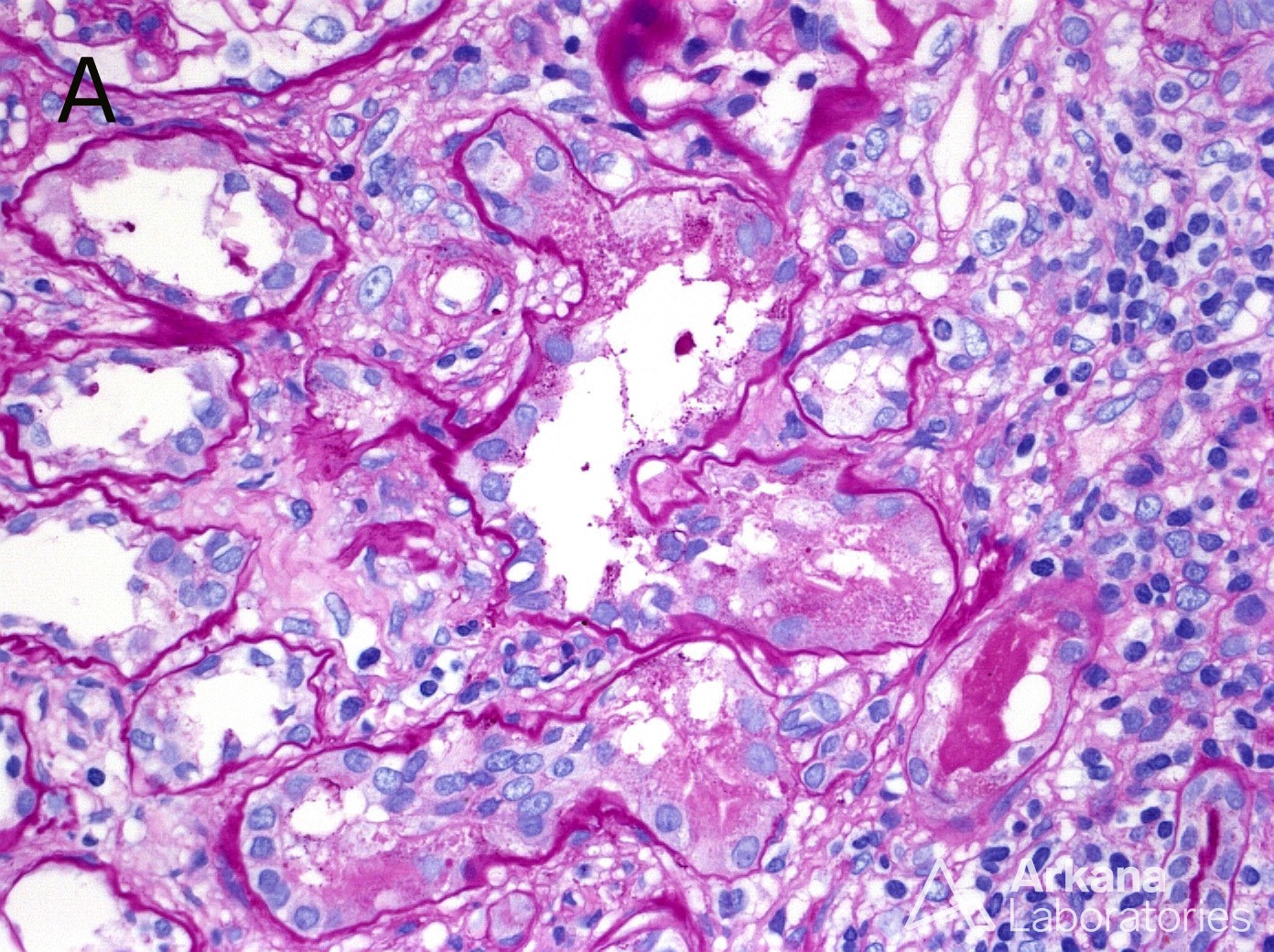
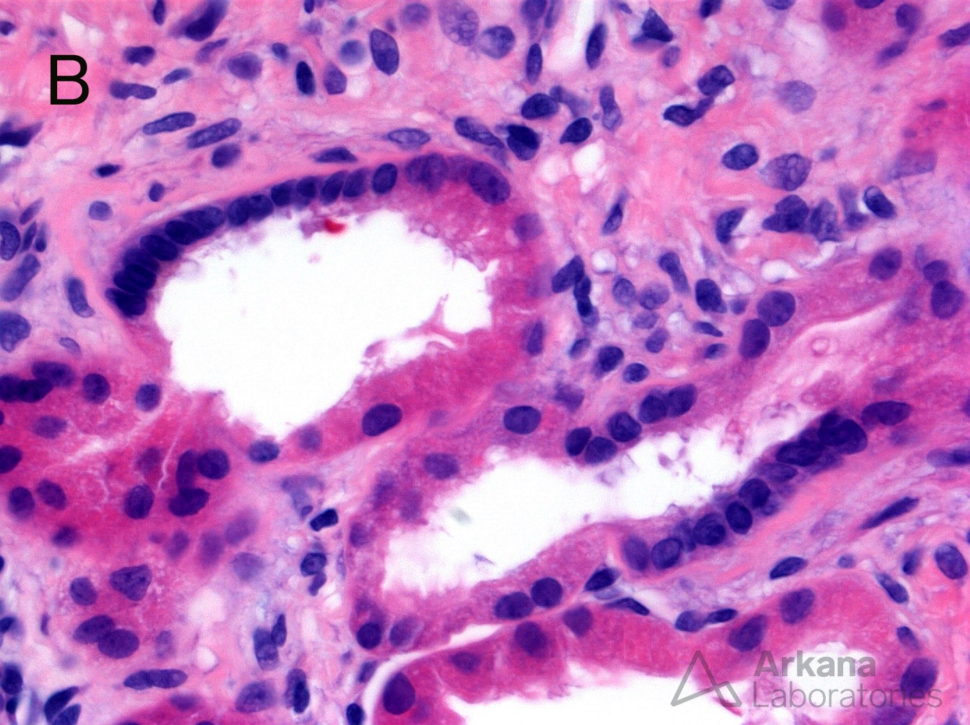
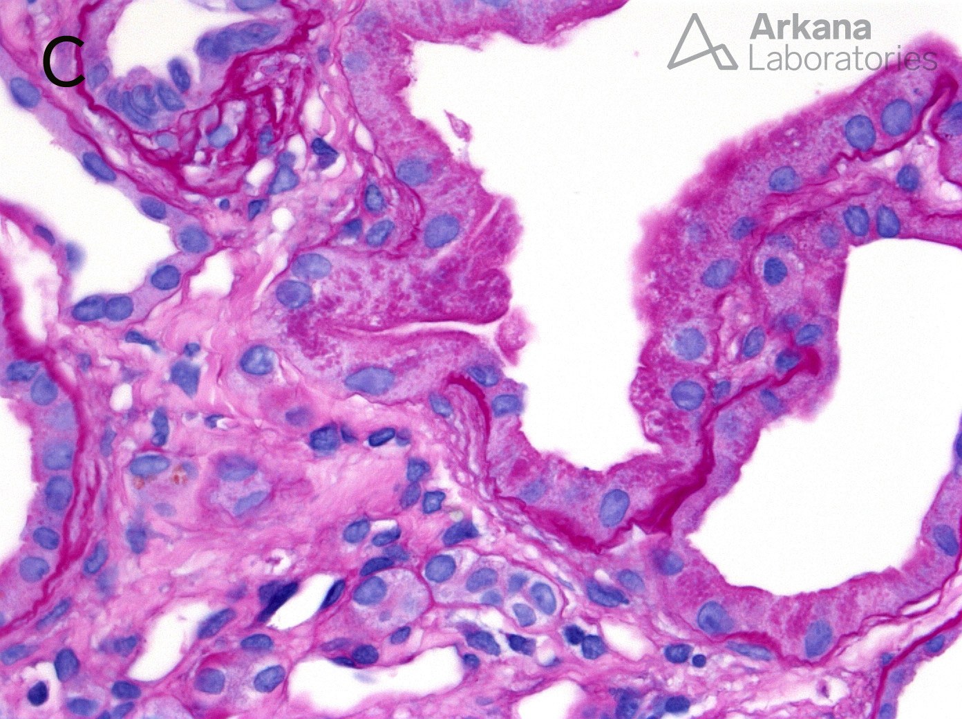
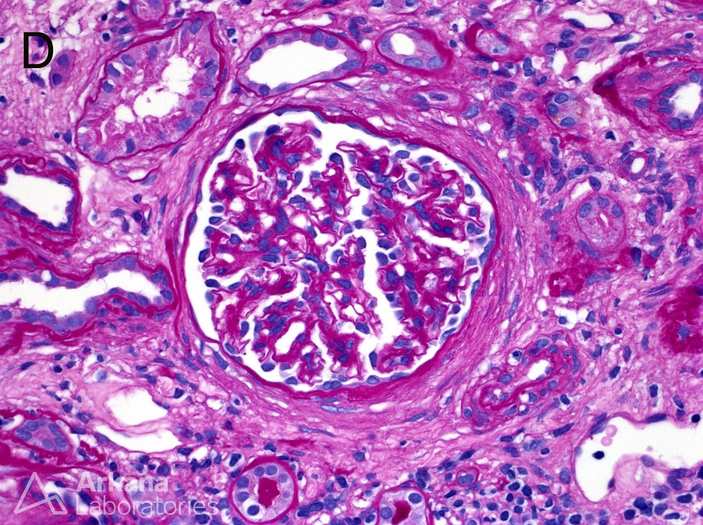
Nephronophthisis is an autosomal recessive tubulointerstitial nephropathy that is a leading genetic etiology of end-stage renal disease in children and young adults. Approximately 60% of patients with a known genetic etiology of nephronophthisis are due to homozygous deletion of the NPHP1 gene. The histopathologic lesions associated with nephronophthisis are shown in these photomicrographs. (A) A tubular floret profile with complicated tubular branching in at least 4 directions (PAS; original magnification ×400). (B) Macula densa-like lesions (arrows) are tubular profiles in which there is a transition to a grouping of cells with crowded, hyperchromatic nuclei and a high nuclear: cytoplasmic ratio (H&E; original magnification ×400). (C) A tubular diverticulum (arrow) with perpendicular outpouching of the tubular cytoplasm and associated overlying TBM thinning (PAS; original magnification ×600). (D) Periglomerular fibrosis with PAS-positive thickening surrounding Bowman’s capsule of an intact glomerulus (PAS; original magnification ×400).
Reference: CP Larsen, SM Bonsib, ML Beggs, JD Wilson. Fluorescence in situ hybridization for the diagnosis of NPHP1 deletion-related nephronophthisis on renal biopsy. 2018. Hum Path. [epub ahead of print]
Quick note: This post is to be used for informational purposes only and does not constitute medical or health advice. Each person should consult their own doctor with respect to matters referenced. Arkana Laboratories assumes no liability for actions taken in reliance upon the information contained herein.