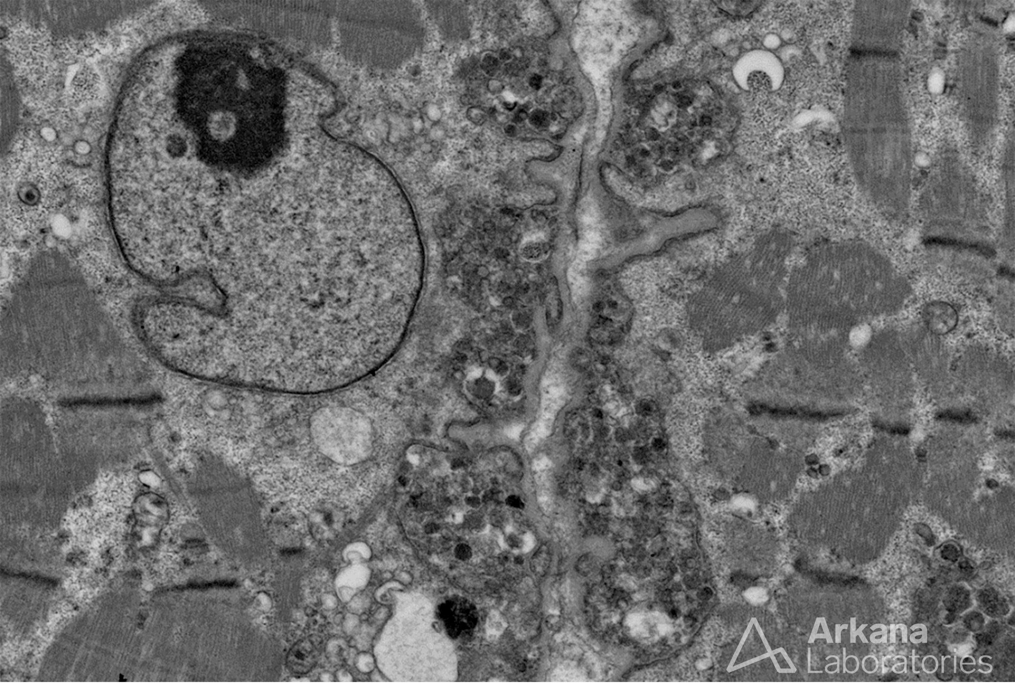
This electron micrograph of two adjacent skeletal muscle cells demonstrates simultaneous exocytosis. Exocytosis in this case shows extravasation of lysosomes from inside the muscle fiber’s cytoplasm by fusion with the plasma membrane (aka sarcolemma) and being expelled into the extracellular space beneath the basement membrane (aka basal lamina). Exocytosis is one mechanism for remodeling of the sarcolemmal membrane that takes place under physiological and pathophysiological conditions, including trafficking of transmembrane machinery, muscle fiber regeneration following injury, and denervation. Much has been written on this topic, but the details at the molecular level are still being elucidated. (Electron micrograph original magnification: 3000x)
Quick note: This post is to be used for informational purposes only and does not constitute medical or health advice. Each person should consult their own doctor with respect to matters referenced. Arkana Laboratories assumes no liability for actions taken in reliance upon the information contained herein.