Peripheral Nerve Axons
This 60-year-old patient underwent sural nerve biopsy to evaluate for neuropathy.
Do the numerous very small punctate areas of immunohistochemical staining arranged in groups seen in Figure #1 correspond to electron microscopy seen in Figure #2 versus Figure #3?
A. Figure #2
B. Figure #3
C. Neither
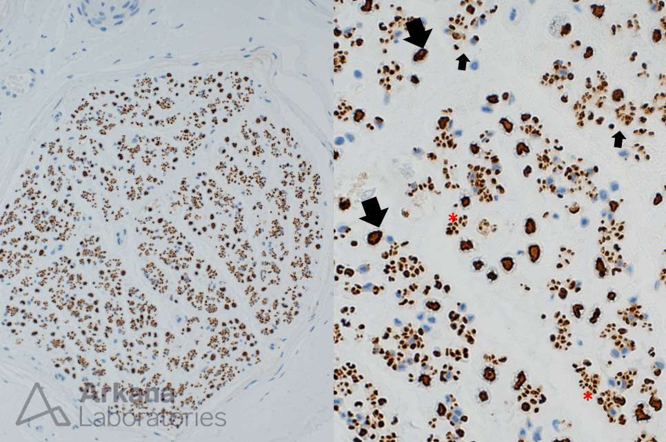
Figure 1: Neurofilament stain FFPE 200x and 600x original magnification
Low (left) and high (right) magnification images showing individual large diameter myelinated axons (large arrows), small diameter myelinated axons (small arrows) and clusters of small diameter unmyelinated axons (red asterix).
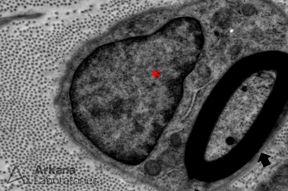
Figure 2: Electron microscopy 2500x magnification
Ultrastructural image showing a single myelinated axon (large arrow) and associated single Schwann cell (nucleus marked by asterix).
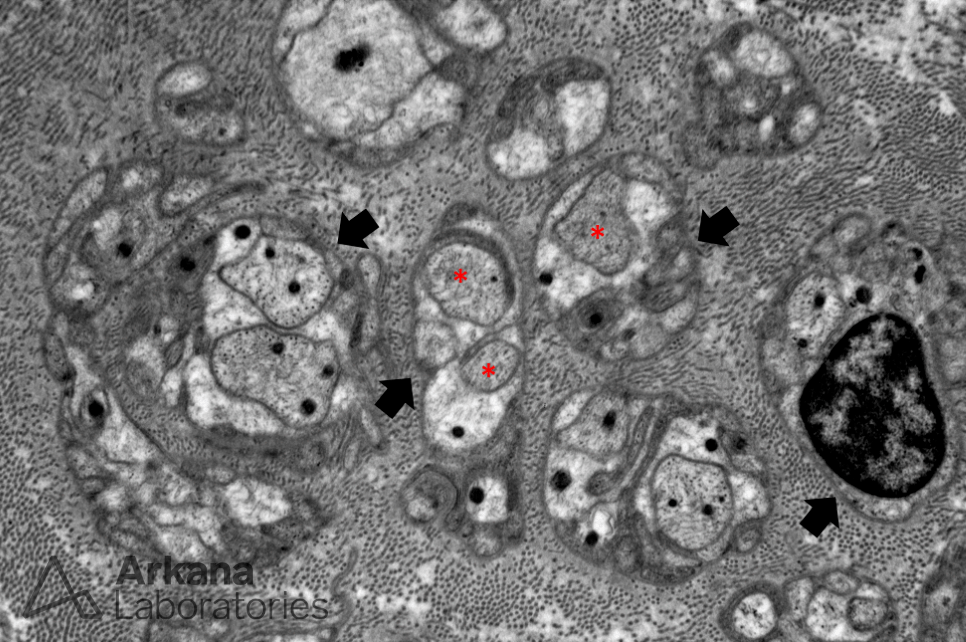
Figure 3: Electron microscopy 4000x magnification
Ultrastructural image showing multiple Schwann cell subunits with one or two associated small diameter unmyelinated axons.
Each Schwann cell subunit is surrounded by basement membrane.
Note the background collagen fibrils.
Answer:
Do the numerous very small punctate areas of immunohistochemical staining arranged in groups seen in Figure #1 correspond to electron microscopy seen in Figure #2 versus Figure #3?
A. Figure #2
B. Figure #3 <– correct answer
C. Neither
Explanation:
The small punctate areas of staining shows in Figure #1 represent neurofilament staining of the small diameter unmyelinated axons. This correlates with the unmyelinated axons seen on electron microscopy in Figure #3. Schwann cells associated with unmyelinated axons in a peripheral nerve may enfold (but not myelinate) multiple axons. In contrast, each myelinated segment of an axon within a peripheral nerve is associated with a single Schwann cell. A myelinated axon is demonstrated in Figure #2.
Note on the neurofilament stain that the number of small diameter unmyelinated axons outnumbers myelinated axons.
Quick note: This post is to be used for informational purposes only and does not constitute medical or health advice. Each person should consult their own doctor with respect to matters referenced. Arkana Laboratories assumes no liability for actions taken in reliance upon the information contained herein.