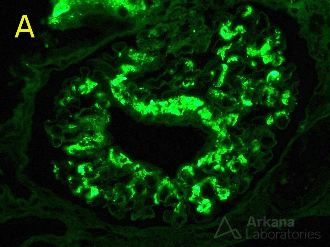
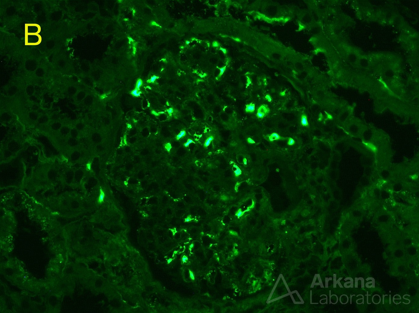
The paraffin immunofluorescence technique is simple to perform for renal pathology laboratories familiar with routine IF. It is important as a salvage technique as well as to identify “masked” deposits. While the interpretation is similar to routine IF, there are important pitfalls to be aware of. Due to fixation, the glomerular capillaries will often contain residual serum on paraffin IF which is not typically present in routine IF sections from frozen tissue. This serum will stain nonspecifically positive for many of the antibodies employed. Photomicrograph A shows strong staining for IgA in the mesangium in a case of IgA nephropathy. However, IgA staining is strongly positive only within the capillary spaces of a glomerulus by paraffin immunofluorescence in photomicrograph B. Since the staining is restricted to the capillary space, this should be interpreted as negative. Identifying the location of the positive staining is the key to avoid misdiagnosing this artifact as positive staining.
Quick note: This post is to be used for informational purposes only and does not constitute medical or health advice. Each person should consult their own doctor with respect to matters referenced. Arkana Laboratories assumes no liability for actions taken in reliance upon the information contained herein.