Clinical History:
This 55-year-old patient presented with complaints of a several year history of slowly progressive asymmetric upper and lower extremity muscle weakness. They also reported that recently they frequently drop objects due to decreased grip strength. CPK level was within normal limits. Myositis specific autoantibody panel and anti-NT5c1A antibody test results were negative. Muscle biopsy was performed to evaluate for myopathy. Only formalin-fixed tissue is available for evaluation.
Tissue was processed for paraffin block embedding. A portion of tissue was post-fixed in glutaraldehyde for possible electron microscopy.
What is your diagnosis based on the images of FFPE H&E stained slide and Toluidine blue stained thick section? What additional testing would you pursue on the tissue available for evaluation to prove this diagnosis?
Answer:
Sporadic Inclusion Body Myositis (sIBM)
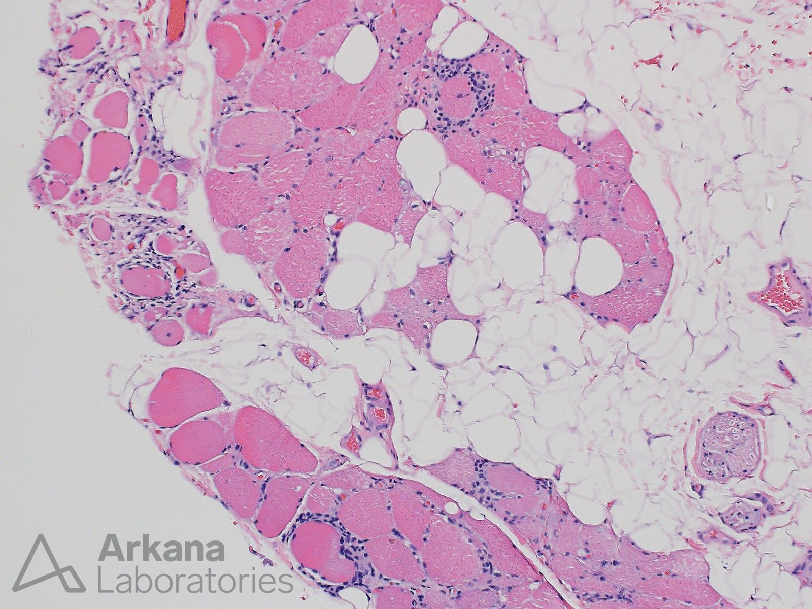
- The FFPE H&E shows features of a chronic active inflammatory myopathic process with partial replacement of skeletal muscle by adipose. Rimmed vacuoles are difficult to impossible to visualize on FFPE sections. In addition, congophilic inclusions seem to be much easier to visualize in frozen sections than FFPE sections.
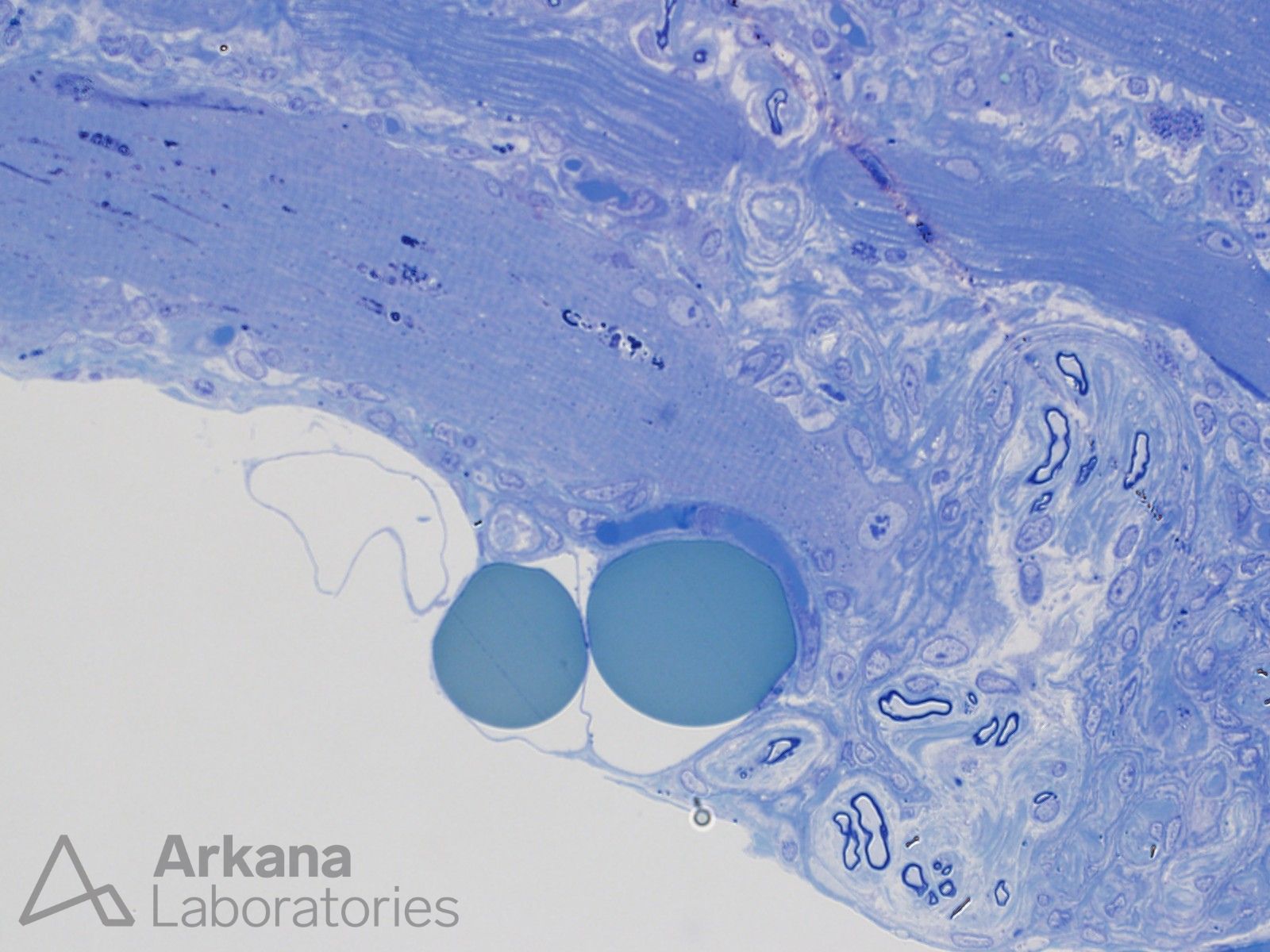
- The Toluidine Blue stained thick section shows a muscle fiber with multiple autophagic vacuole type structures. Note the adjacent unremarkable small intramuscular nerve twigs containing myelinated axons.
Additional Testing:
- IHC for TDP43 and p62 to evaluate for protein aggregates
- Electron microscopy to evaluate for sarcoplasmic or intranuclear 15-21 nm thick amyloid-like fibrillary inclusions
Reference(s) / Additional Reading:
- Guglielmi V, Cheli M, Tonin P, Vattemi G. Sporadic Inclusion Body Myositis at the Crossroads between Muscle Degeneration, Inflammation, and Aging. Int J Mol Sci. 2024 Feb 27;25(5):2742. doi: 10.3390/ijms25052742. PMID: 38473988; PMCID: PMC10932328.
- Weihl CC, Mammen AL. Sporadic inclusion body myositis – a myodegenerative disease or an inflammatory myopathy. Neuropathol Appl Neurobiol. 2017 Feb;43(1):82-91. doi: 10.1111/nan.12384. PMID: 28111778.
- Lilleker JB, Naddaf E, Saris CGJ, Schmidt J, de Visser M, Weihl CC; 272nd ENMC workshop participants. 272nd ENMC international workshop: 10 Years of progress – revision of the ENMC 2013 diagnostic criteria for inclusion body myositis and clinical trial readiness. 16-18 June 2023, Hoofddorp, The Netherlands. Neuromuscul Disord. 2024 Apr;37:36-51. doi: 10.1016/j.nmd.2024.03.001. Epub 2024 Mar 7. PMID: 38522330.
- Greenberg SA. Inclusion body myositis: clinical features and pathogenesis. Nat Rev Rheumatol. 2019 May;15(5):257-272. doi: 10.1038/s41584-019-0186-x. PMID: 30837708.
Quick note: This post is to be used for informational purposes only and does not constitute medical or health advice. Each person should consult their own doctor with respect to matters referenced. Arkana Laboratories assumes no liability for actions taken in reliance upon the information contained herein.