This 40-year-old patient presented with complaints of leg cramps, lower extremity muscle weakness, and loss of muscle mass. Laboratory studies demonstrated mild elevation of CPK (475). Physical examination demonstrated proximal and distal muscle weakness involving both lower extremities.
Which of the following accounts for the circular areas of dark staining with surrounding zone of pale staining on the modified Gomori Trichrome stained section?
A. Target fibers
B. Rimmed vacuoles
C. Centronuclear myopathy
D. Cytoplasmic bodies
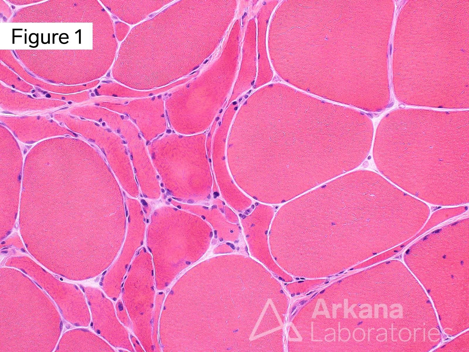
Hematoxylin and eosin stained section shows a small group of acutely angulated atrophic muscle fibers (grouped atrophy).
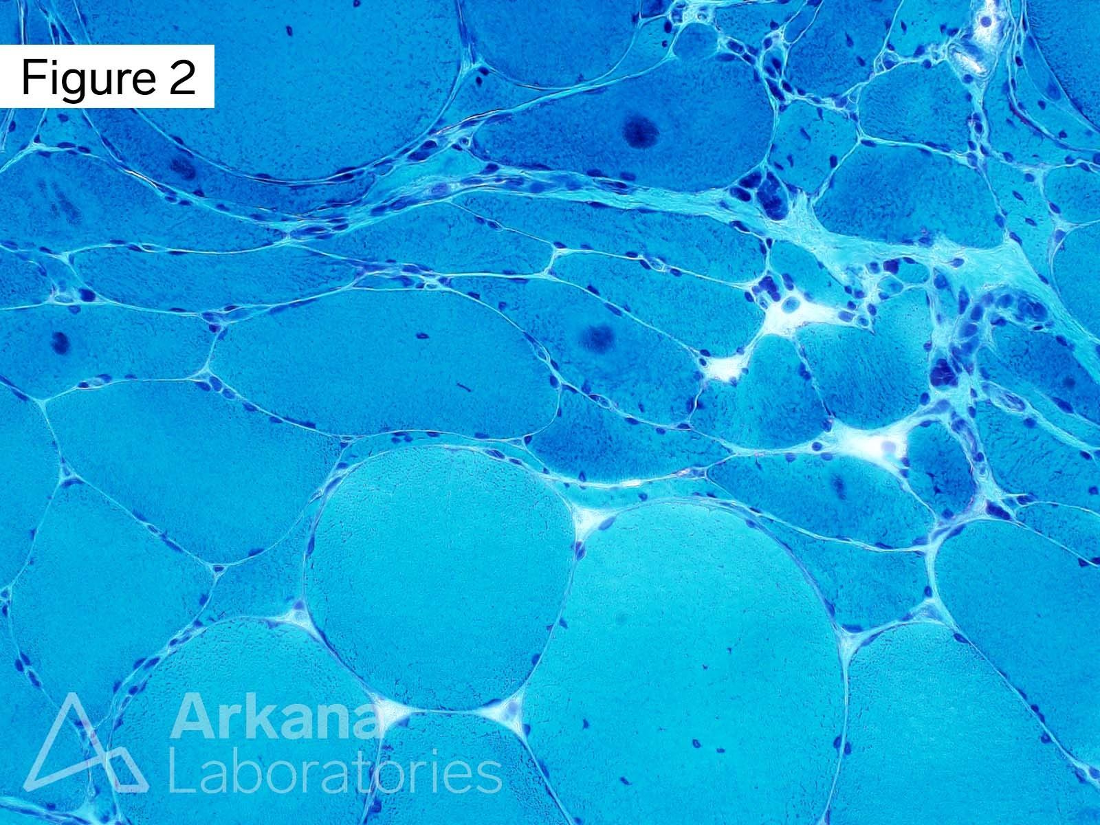
Modified Gomori Trichrome stained section shows multiple acutely angulated atrophic muscle fibers with round central to eccentric areas of darker staining and a subtle rim of surrounding pale staining (for examples see arrows). Note that these areas disrupt the normal lace-like intermyofibrillar mitochondrial staining in the remainder of the involved muscle fibers. These areas are visible on the H&E stained section (see figure 1) but are less obvious. This highlights the utility of special stains that bring out features that may not be well-demonstrated on H&E.
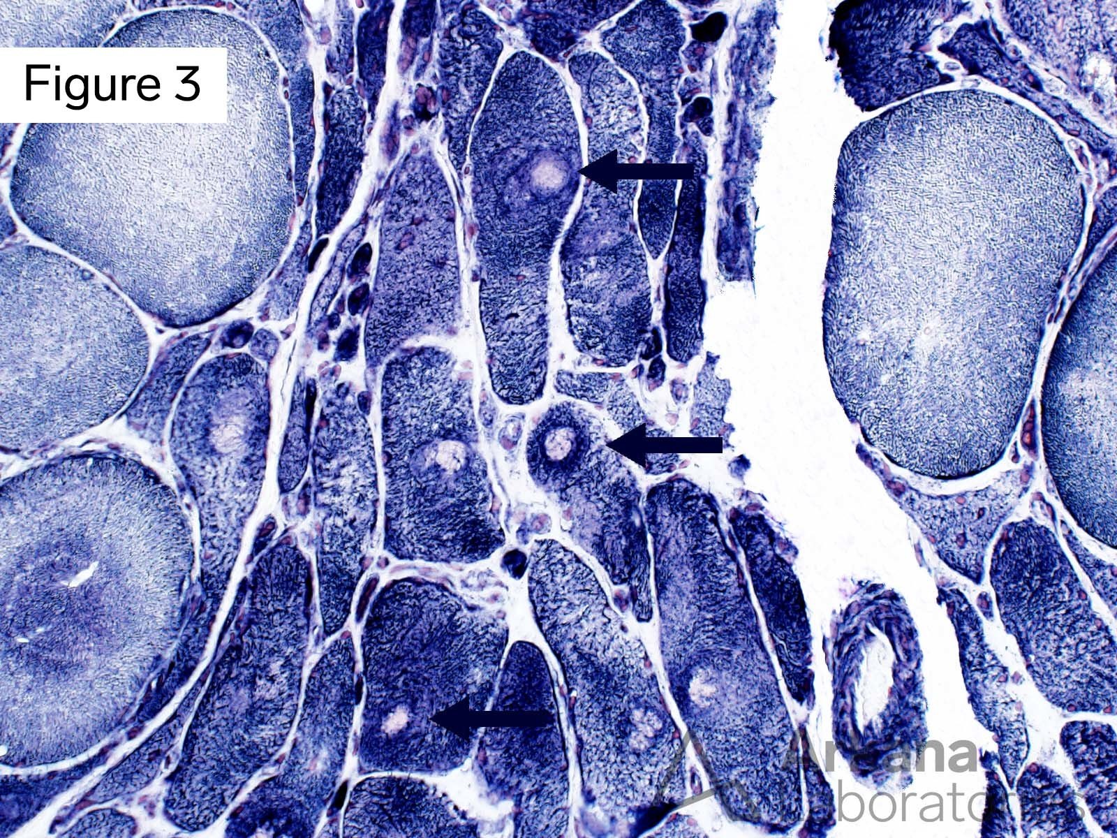
Nicotinamide Adenine dinucleotide Dehydrogenase-Tetrazolium Reductase (NADH) stained section demonstrates that the areas seen on modified Gomori Trichrome show pale central staining with a surrounding area of dark staining, and retained normal staining in the remainder of the muscle fibers consistent with “target fibers” (see arrows for examples). Target fibers are most commonly seen in the setting of denervation.
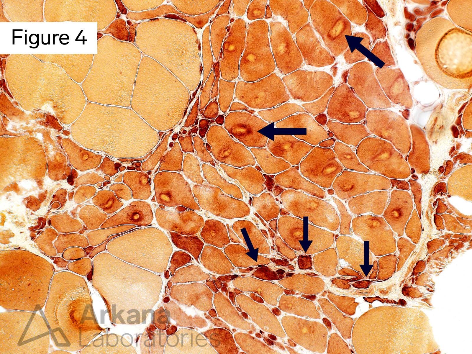
In this case, the target fibers are also seen on the Esterase stained section (see big arrows). Esterase stain also shows diffuse increased sarcoplasmic staining of occasional angulated atrophic fibers (see thinner arrows). Normally, esterase staining is limited to neuromuscular junctions where the enzyme cholinesterase is located. Esterase positive acutely angulated fibers are another common feature seen in the setting of denervation.
Answer: Target fibers
Target fibers are most commonly seen in the setting of denervation (the diagnosis in this case). While they are best demonstrated on NADH-TR preparation, they are visible with other stains.
Along with H&E, modified Gomori Trichrome is an excellent stain for the general morphologic evaluation of skeletal muscle frozen sections. It highlights mitochondria (for example “ragged-red muscle fibers”) and a variety of inclusions (nemaline rods, cytoplasmic bodies, tubular aggregates, and other). Because it highlights the lace-like pattern of mitochondria between the bundles of sarcomeres in normal muscle, it is useful for detecting disorganization of myofibrillar arrangement in which there is associated redistribution of mitochondria. Given its utility, some experts in the pathologic evaluation of skeletal muscle biopsies evaluate the modified Gomori Trichrome section before taking a look at the H&E.
The staining characteristics demonstrated in figure 2 are not those of rimmed vacuoles, muscle fiber nuclei, or cytoplasmic bodies.
References
Engel WK. Muscle target fibres, a newly recognized sign of denervation. Nature. 1961 Jul 22;191:389-90. doi: 10.1038/191389a0. PMID: 13696814.
Schmitt HP, Volk B. The relationship between target, targetoid, and targetoid/core fibers in severe neurogenic muscular atrophy. J Neurol. 1975 Sep 22;210(3):167-81. doi: 10.1007/BF00316244. PMID: 51074.
Schmitt HP, Volk B. The relationship between target, targetoid, and targetoid/core fibers in severe neurogenic muscular atrophy. J Neurol. 1975 Sep 22;2
Schmitt HP, Volk B. The relationship between target, targetoid, and targetoid/core fibers in severe neurogenic muscular atrophy. J Neurol. 1975 Sep 22;210(3):167-81. doi: 10.1007/BF00316244. PMID: 51074.10(3):167-81. doi: 10.1007/BF00316244. PMID: 51074.
Quick note: This post is to be used for informational purposes only and does not constitute medical or health advice. Each person should consult their own doctor with respect to matters referenced. Arkana Laboratories assumes no liability for actions taken in reliance upon the information contained herein.
