Happy New Year from the Neuro Notes team!
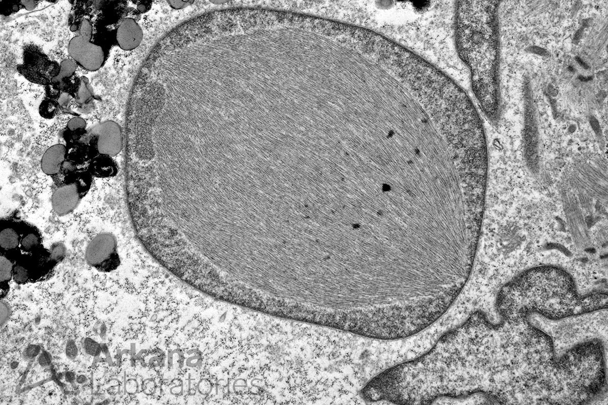
This diagnostic electron microscopy image shows an amyloid-like intranuclear inclusion in a patient with sporadic Inclusion Body Myositis (sIBM). Notice the intrasarcoplasmic lipofuscin pigment toward the left middle and upper region of the image and the disorganized sarcomeres toward the right middle and upper region of the image.
Quick note: This post is to be used for informational purposes only and does not constitute medical or health advice. Each person should consult their own doctor with respect to matters referenced. Arkana Laboratories assumes no liability for actions taken in reliance upon the information contained herein.
