Clinical History:
This 76-year-old male presented with complaints of muscle pain. Their past medical history is significant for Sjogren’s syndrome, fibromyalgia, peripheral neuropathy, hypertension and hyperlipidemia. Laboratory studies showed normal CPK and elevated CRP. No myotoxic medications were noted in the patient’s home medication list.
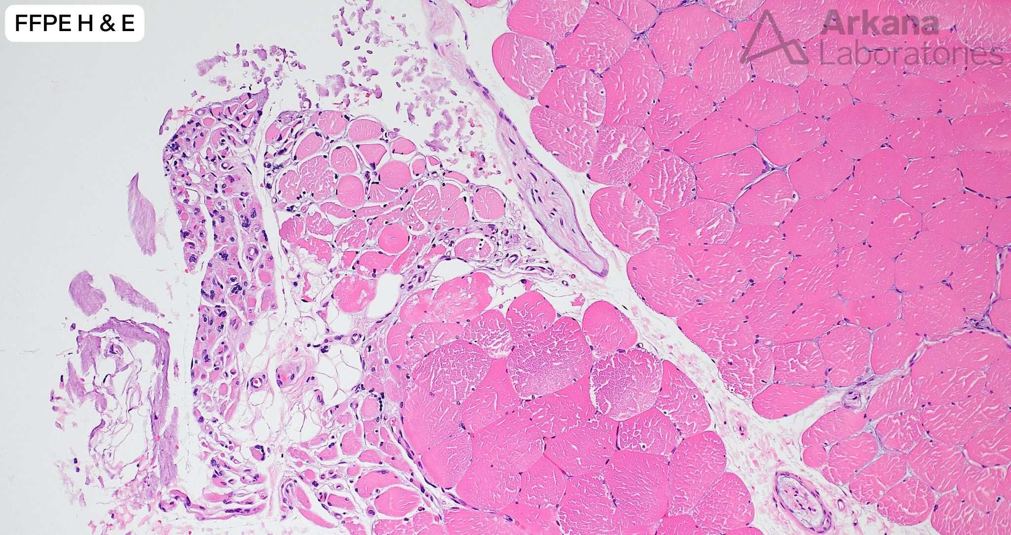
What are your diagnoses for the changes seen in the muscle biopsy (image #1) and the incidental finding that was seen in multiple blood vessels (images #2 and #3)?
A. Denervation
B. Denervation, vascular amyloid
C. Denervation, arteriosclerosis
D. Inflammatory myopathy


Answer:
Denervation (neurogenic atrophy) and incidental vascular amyloid deposition
- The muscle biopsy showed well-developed grouped atrophy and hypertrophy, most consistent with denervation type changes. Enzyme histochemical stains further confirmed this impression; frequent esterase positive atrophic muscle fibers indicating the presence of ongoing denervation, and fiber type grouping on immunohistochemical stains for developmental heavy chain (fast and slow isoforms) indicating prior denervation and subsequent successful reinnervation.
- No inflammation or morphologic features of inflammatory myopathy are seen. It must be noted that variable numbers of regenerating myofibers may be seen in the setting of denervation (felt to represent myofiber injury related to continued use of a partially denervated muscle).
- Occasional blood vessels showed variably sized areas of somewhat hyalin appearing material within the blood vessel wall. Congo red stain shows that this represents amyloid type material.
- The appearance of amyloid deposition is different than what would be expected for arteriosclerosis; and arteriosclerosis would not stain for Congo Red.
- Amyloidosis can be associated with peripheral neuropathy. In this case, no amyloid was noted in multiple intramuscular nerve twigs that were present in the biopsy sample.
Reference(s) / Additional Reading:
- Pinto MV, Dyck PJB, Liewluck T. Neuromuscular amyloidosis: Unmasking the master of disguise. Muscle Nerve. 2021 Jul;64(1):23-36. doi: 10.1002/mus.27150. Epub 2021 Jan 17. PMID: 33458861.
Quick note: This post is to be used for informational purposes only and does not constitute medical or health advice. Each person should consult their own doctor with respect to matters referenced. Arkana Laboratories assumes no liability for actions taken in reliance upon the information contained herein.