Clinical History:
This 25-year-old patient presented with a one-year history of progressive bilateral upper and lower extremity muscle weakness and painful paresthesias. Laboratory testing showed vitamin B12 and folate deficiency. Electrodiagnostic studies showed features of a sensorimotor neuropathy with decreased amplitude and velocity (mixed axonopathic and demyelinating). Nerve biopsy was performed in an attempt to further determine the etiology of this patient’s neuropathy.
What abnormality is demonstrated on this electron microscopic image?
A. Degenerating axon
B. Regenerated cluster
C. Active demyelination
D. Normal nerve
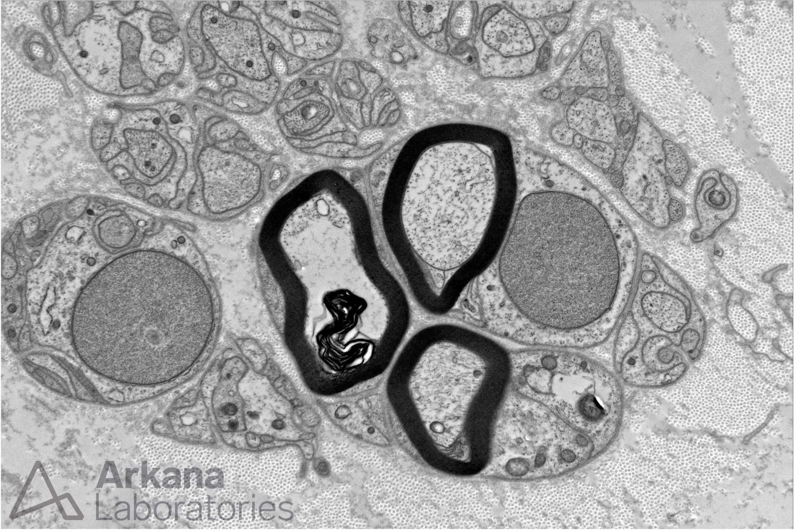
Answer:
Regenerated Cluster
- The image shows a cluster of three thinly myelinated large diameter axons that are too close together. Regenerated clusters are evidence of prior axonal injury with subsequent recovery through stages of early axonal sprout formation and later remyelination.
- Note the adjacent non-myelinating Schwann cell subunits and their associated small diameter non-myelinated axons.
- Note the basement membrane surrounding the Schwann cells.
- Degenerating axons show myelin debris without a visible intact axon, while axons undergoing demyelination show myelin debris and/or stripping of myelin from intact axons by macrophages with a visible intact axon.
Reference(s) / Additional Reading:
- Please see prior NeuroNotes from April 11, 2024.
Quick note: This post is to be used for informational purposes only and does not constitute medical or health advice. Each person should consult their own doctor with respect to matters referenced. Arkana Laboratories assumes no liability for actions taken in reliance upon the information contained herein.