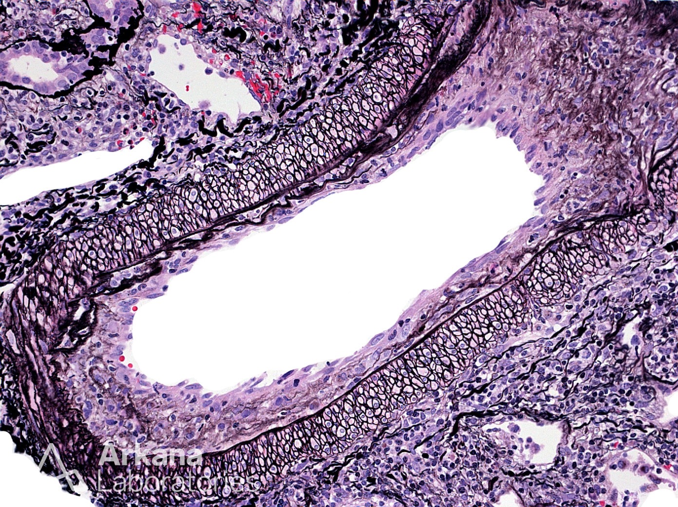
What is this finding and what is the most likely etiology?
This is a Jones silver stain showing intimal arteritis. The endothelial, intimal vascular layer shows reactive changes and is pushed upward into the lumen by the presence of numerous subintimal inflammatory cells (mostly lymphocytes). This finding is most commonly seen in the transplant setting where it represents a rejection process and is graded by the amount of lumen compromise noted. While traditionally thought to represent an acute T-cell mediated rejection process, we now know that intimal arteritis can also occur from antibody mediated rejection as well. As both etiologies have identical morphology, segregation into T-cell mediated or antibody-mediated requires evaluation of the glomeruli and tubulointerstitium for other supporting morphologies. In rare cases, the presence of an isolated vascular rejection or a mixed T-cell and antibody mediated rejection can make this segregation impossible by morphology alone. In the native kidney intimal arteritis is a very uncommon finding. But, in our experience it is usually secondary to cryoglobulinemia when present.
Quick note: This post is to be used for informational purposes only and does not constitute medical or health advice. Each person should consult their own doctor with respect to matters referenced. Arkana Laboratories assumes no liability for actions taken in reliance upon the information contained herein.