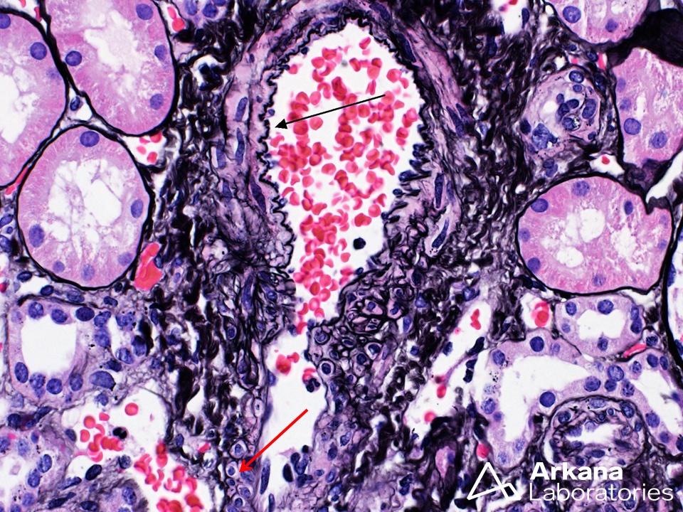
The image captures the transition of an arteriole (arteriole transition) as it loses its internal elastic lamina (black arrow). Note that the smaller arteriole branch, while lacking this elastic layer, still retains smooth muscle cells (red arrow). The elastic tissue adds structural integrity to arterial and larger arteriolar walls during pulsatile distension from higher intravascular pressure, and the retained muscular layer in small arterioles helps provide an important component of the pre-capillary sphincter mechanism, whereby regulation of blood flow to capillary beds occurs.
Quick note: This post is to be used for informational purposes only and does not constitute medical or health advice. Each person should consult their own doctor with respect to matters referenced. Arkana Laboratories assumes no liability for actions taken in reliance upon the information contained herein.