Search & Filter All Posts
Results for Diagnose This!
(159 Results)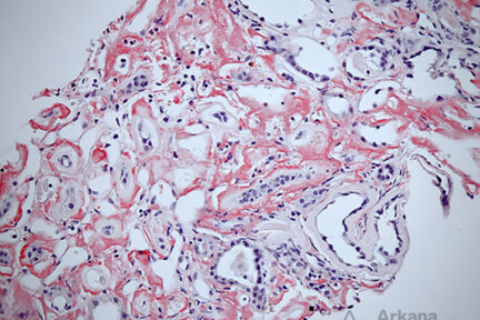
Diagnose This 4/29/2024
What would be your leading Dx and subtype based on the Congo red image above showing deposition predominately in the…
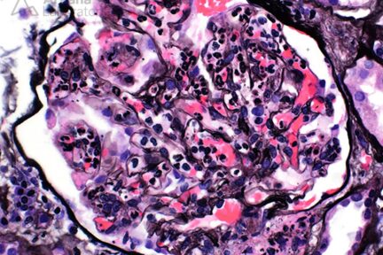
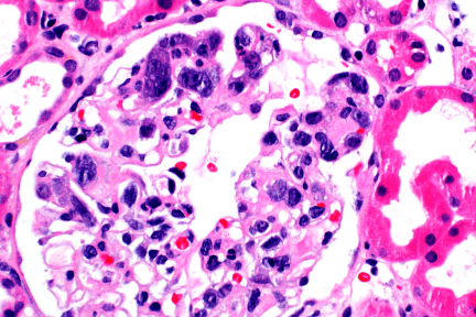
Diagnose This 4/15/2024
Based on the image provided, what would be your leading diagnosis with C3-dominant immune-complex deposition? The light microscopic image depicts…
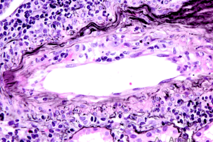
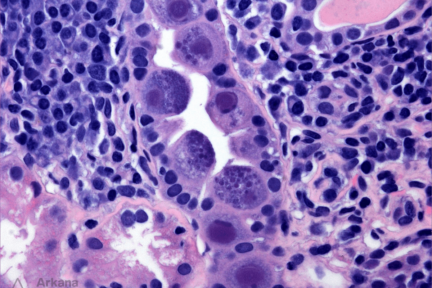
Diagnose This 4/1/2024
What is this finding and what is the significance in the transplant setting? The light microscopic image depicts an…
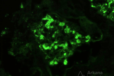
Diagnose This 3/18/2024
What is at the top of your differential in a patient with a positive SPEP (IgG kappa) and the immunofluorescence…
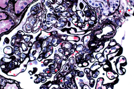
Diagnose This 3/11/2024
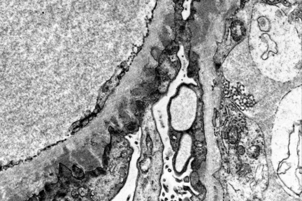
What is your diagnosis in a patient with nephrotic syndrome? The electron photomicrograph demonstrates frequent subepithelial deposits along the glomerular…
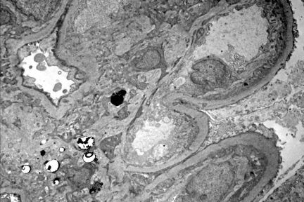
Diagnose This 3/4/2024
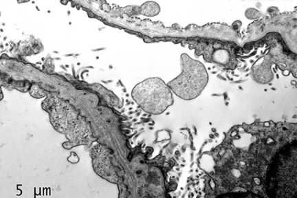
The ultrastructural abnormalities present in the image are suspicious for what underlying disease? The electron photomicrograph demonstrates two abnormal…
Diagnose This 2/26/2024
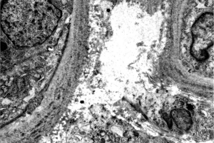
What is your diagnosis based on the EM image provided? The electron microscopy image provided is that of portions of…
Diagnose This 2/19/2024
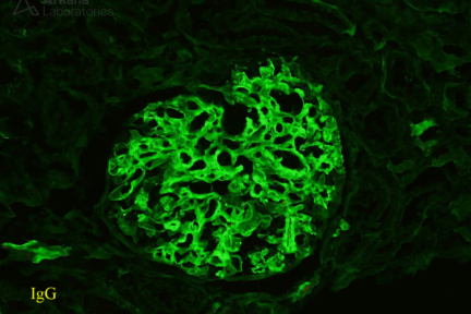
What is your leading diagnosis when seeing this particular IgG staining pattern? Answer: Fibrillary Glomerulopathy The immunofluorescence shows…
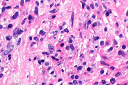
Diagnose This 2/12/2024
What is your diagnosis? The light microscopic image depicts a renal transplant biopsy with multiple histiocytes containing intracytoplasmic, targetoid…
Diagnose This 2/5/2024
What is your diagnosis in this transplant biopsy given the features present? Answer: Chronic active antibody-mediated rejection The features…
Diagnose This 1/29/2024
What stain would you order to clinch the diagnosis in this transplant patient with AKI? A CMV stain is…
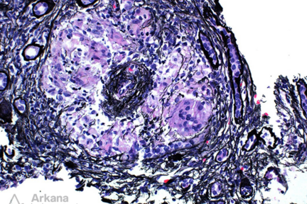
Diagnosis This 1/15/2024
What considerations should be included in your differential diagnosis given the provided histologic finding? The image of a Jones silver…
Diagnose This! (January 9, 2023)
This is the biopsy of a 45 yo with alcoholic cirrhosis, bacteremia, hematuria, proteinuria and AKI. IF shows IgA (3+),…
Diagnose This (October 10, 2022)
This is the biopsy of a 47-year-old female with cervical cancer, on anti-VEGF therapy, with proteinuria and normal renal function.…
Diagnose This (September 26, 2022)
This is the biopsy from a 73-year-old with AKI and normal bone marrow biopsy done for investigation of anemia and…