Search & Filter All Posts
Results for eyeSCANdy
(86 Results)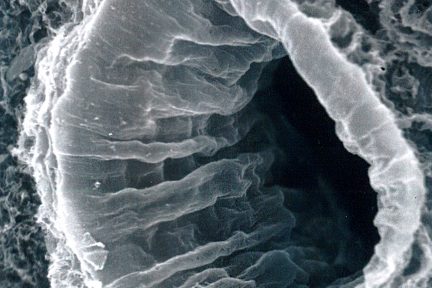
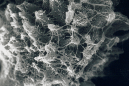
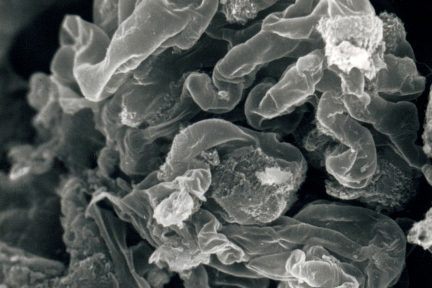
Corrugated Lumen of a Renal Artery
Today’s eyeSCANdy image shows corrugated lumen of a renal artery following removal of endothelial cells!
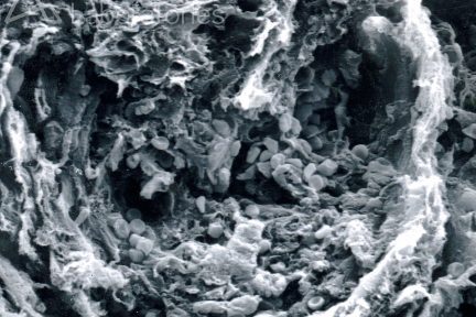
Acellular Scanning EM with Fibrocellular Crescent
This eyeSCANdy image shows acellular scanning EM with a fibrocellular crescent with entrapped RBCs. The glomerulus is not present.
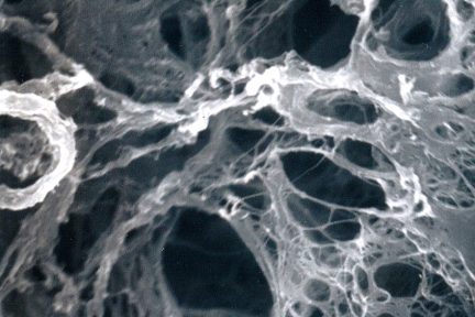
Acellular Scanning EM with Empty Lacunar Spaces
Todays eyeSCANdy image shows acellular scanning EM with empty lacunar spaces of a fibrocellular crescent previously occupied by cells.
Segmental Necrosis
Todays eyeSCANdy image shows acellular scanning EM with normal glomerular tuft at top and segmental necrosis at bottom.…
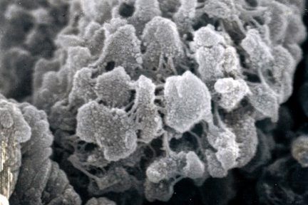
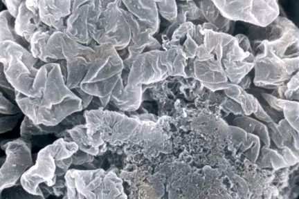
Spicular Amyloid Deposit Showing Blunt Conical Formations
This eyeSCANdy image shows spicular amyloid deposit showing blunt conical formations with interconnecting fibrils.
Glomerular Tuft with Intact Architecture
Today’s eyeSCANdy image shows glomerular tuft with overall intact architecture. Discrete masses of specular amyloid (long arrows) interrupt smooth uninvolved…
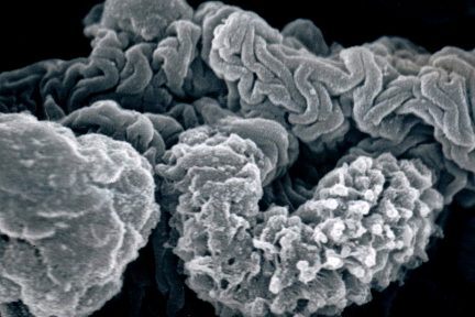
Glomerular Tuft with Collapsed GBM
This eyeSCANdy image shows portions of glomerular tuft with collapsed GBM (across top), spicular amyloid (middle right), and nodular amyloid…
Conical Masses Interconnected by Branching Fibrils
This eyeSCANdy image shows portions of glomerular tuft with collapsed GBM (across top), spicular amyloid (middle right), and nodular amyloid…
Glomerular Tuft
Today’s eyeSCANdy image shows glomerular tuft with overall intact architecture. Discrete masses of specular amyloid interrupt smooth uninvolved segments of…
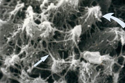
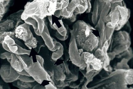
Spicular Amyloid With Tapering Conical Masses
This image shows spicular amyloid with tapering conical masses (curved arrow) interconnected by branching fibrils (arrows).
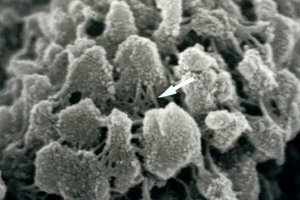
Spicular Amyloid Deposit Showing Blunt Conical Formations
This image shows spicular amyloid deposit showing blunt conical formations with interconnecting fibrils (arrow).