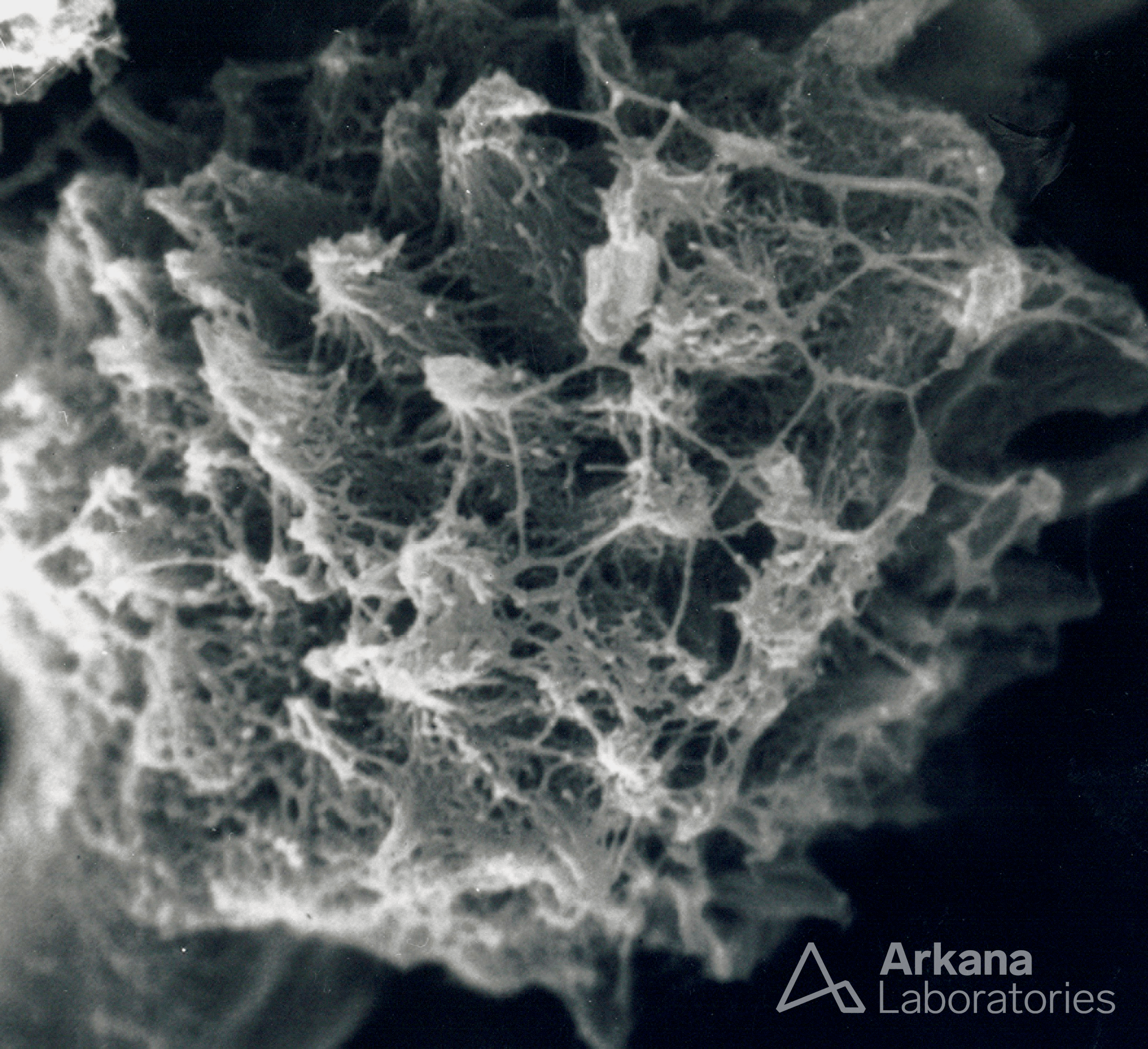
This eyeSCANdy image shows portions of glomerular tuft with collapsed GBM (across top), spicular amyloid (middle right), and nodular amyloid (middle left).
Quick note: This post is to be used for informational purposes only and does not constitute medical or health advice. Each person should consult their own doctor with respect to matters referenced. Arkana Laboratories assumes no liability for actions taken in reliance upon the information contained herein.