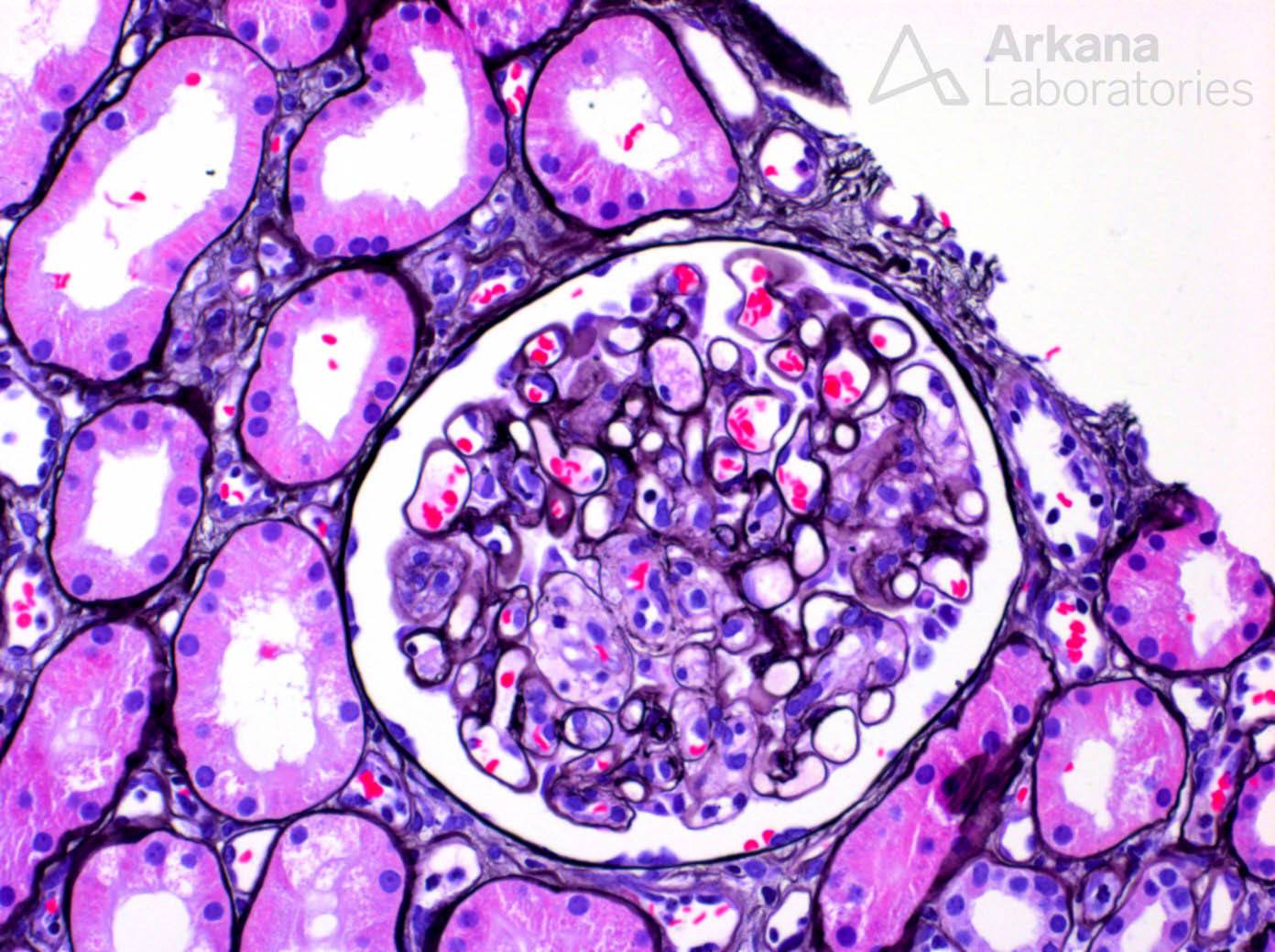
What is this finding in this native kidney?
The photomicrograph shows a glomerulus on Jones silver stain with segmental areas of flocculent material with hypercellularity that is consistent with mesangiolysis. Mesangiolysis can be seen in the setting of several diseases such as diabetic glomerulopathy, thrombotic microangiopathy, proliferative glomerulonephritis, membranoproliferative patterns of injury, calcineurin inhibitor toxicity, and antibody-mediated rejection in the transplant. In this image, no mesangial expansion is present to suggest diabetic glomerulopathy, and no endocapillary proliferation is seen. Additionally, no membranoproliferative pattern of injury is seen. However, this patient did have a history of lung carcinoma, gemcitabine exposure, and bevacizumab exposure all potential etiologies of thrombotic microangiopathy. And, while not shown here, the biopsy showed other, more impressive signs of thrombotic microangiopathy such as glomerular fibrin thrombi, RBC fragments within mesangiolysis, and subendothelial lucent widening of the lamina rara interna on electron microscopy.
Quick note: This post is to be used for informational purposes only and does not constitute medical or health advice. Each person should consult their own doctor with respect to matters referenced. Arkana Laboratories assumes no liability for actions taken in reliance upon the information contained herein.