What is your diagnosis?
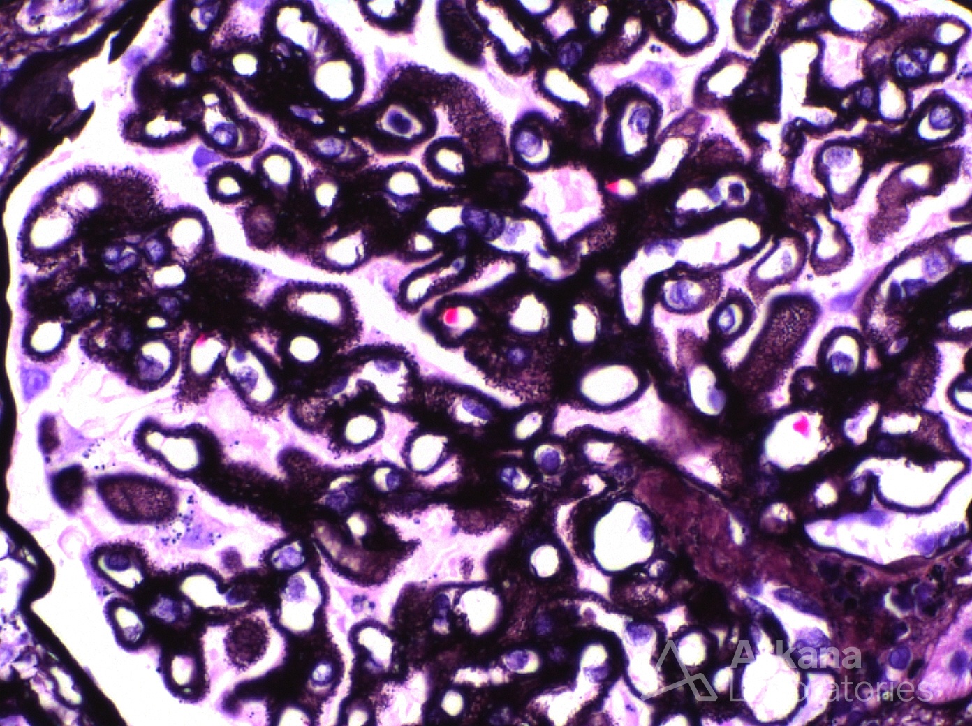
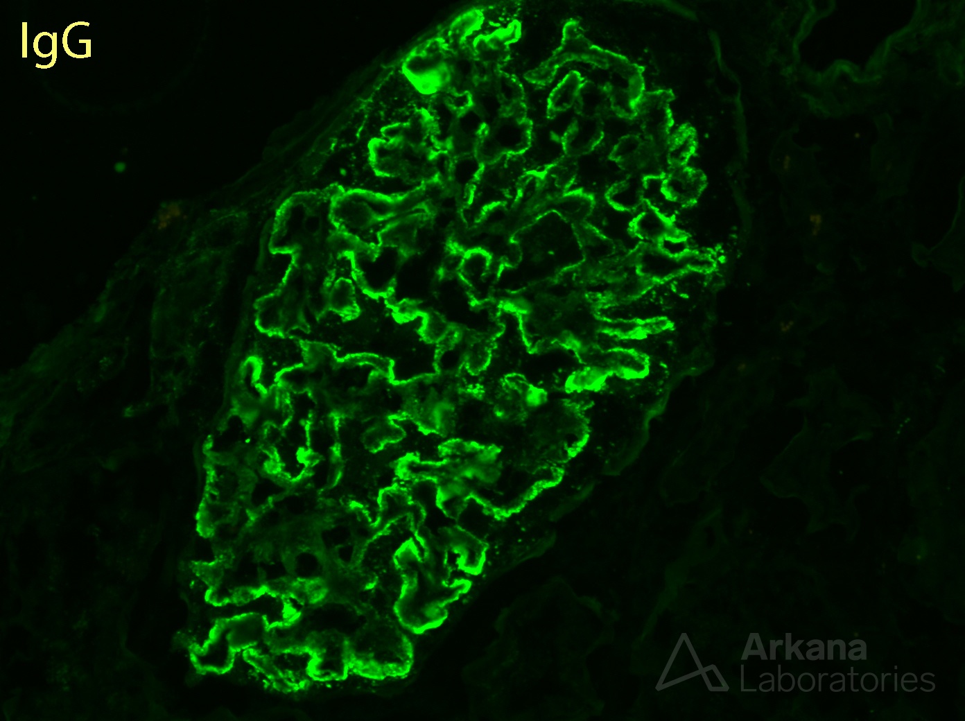
The photomicrograph shows a high power image of a glomerulus on Jones methenamine silver stain. If you look closely you can see numerous, small holes in the glomerular basement membranes as well as extensive spike formation. This is a classic finding of membranous glomerulopathy which is the diagnosis, although it is not exclusive to this disease. Interestingly, while this finding is present in the majority of membranous glomerulopathy cases, hole and spike formation typically represent immune complex deposits that are either intramembranous or deep subepithelial and should the disease be caught early, hole and spike formation may not be seen. However, by immunofluorescence diffuse, granular, capillary wall IgG (see image below) staining is present even in very early cases.
Quick note: This post is to be used for informational purposes only and does not constitute medical or health advice. Each person should consult their own doctor with respect to matters referenced. Arkana Laboratories assumes no liability for actions taken in reliance upon the information contained herein.