In the presence of a negative immunofluorescence panel, what is your diagnosis?
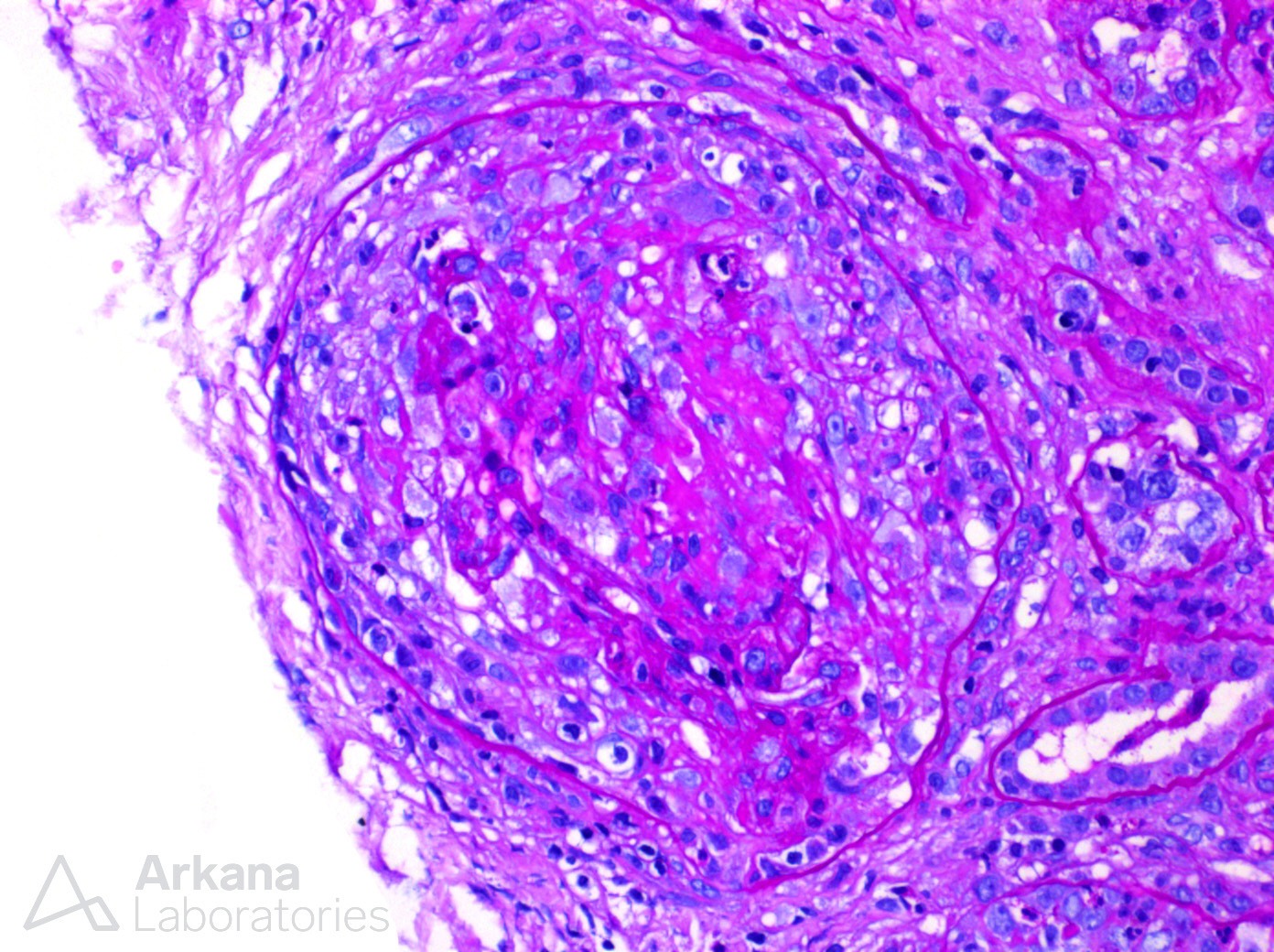
This photomicrograph shows a high power image of a glomerulus with global crescent formation and central tuft fibrinoid necrosis on a PAS stained section. While crescent formation can occur in a variety of kidney diseases, they are typically stratified into three classes: Anti-glomerular basement membrane antibody (AGBM) disease, Pauci-immune type, and immune complex-mediated. These classes are stratified by immunofluorescence with AGBM showing linear IgG staining of the capillary loops and immune complex-mediated showing a pattern of immune complex deposition consistent with the underlying etiology (such as full house staining in lupus nephritis or smudgy IgG staining in fibrillary glomerulopathy). Pauci-immune type shows minimal to no immune complex deposition and is usually associated with ANCA-mediated disease. Of note, endocarditis-associated glomerulonephritis can also present with a pauci-immune and crescentic morphologic pattern and may warrant exclusion in at-risk patients or if clinical concern exists.
Quick note: This post is to be used for informational purposes only and does not constitute medical or health advice. Each person should consult their own doctor with respect to matters referenced. Arkana Laboratories assumes no liability for actions taken in reliance upon the information contained herein.