What is your diagnosis?
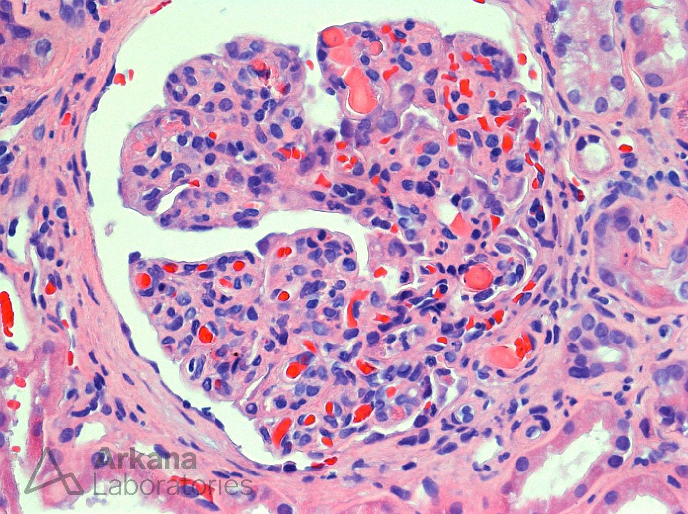
The photomicrograph shows an H&E stain of a hypercellular glomerulus with a lobular architecture most compatible with membranoproliferative pattern glomerulonephritis. At 12- and 4-o’clock there are intracapillary hyaline thrombi present. While not specific, this combination of findings is most concerning for an immune complex mediated glomerulonephritis such as lupus nephritis or cryoglobulinemic glomerulonephritis. However, this patient had no history of autoimmune disease and negative ANA/dsDNA serologies. By immunofluorescence, IgG, IgM, C3, kappa and lambda positivity within the capillary walls and mesangium were noted. Upon further workup the patient was found to be hepatitis C positive. Subsequent studies were positive for serum cryoglobulins and positive rheumatoid factor with a decreased C4 level confirming the diagnosis of cryoglobulinemic glomerulonephritis.
Quick note: This post is to be used for informational purposes only and does not constitute medical or health advice. Each person should consult their own doctor with respect to matters referenced. Arkana Laboratories assumes no liability for actions taken in reliance upon the information contained herein.