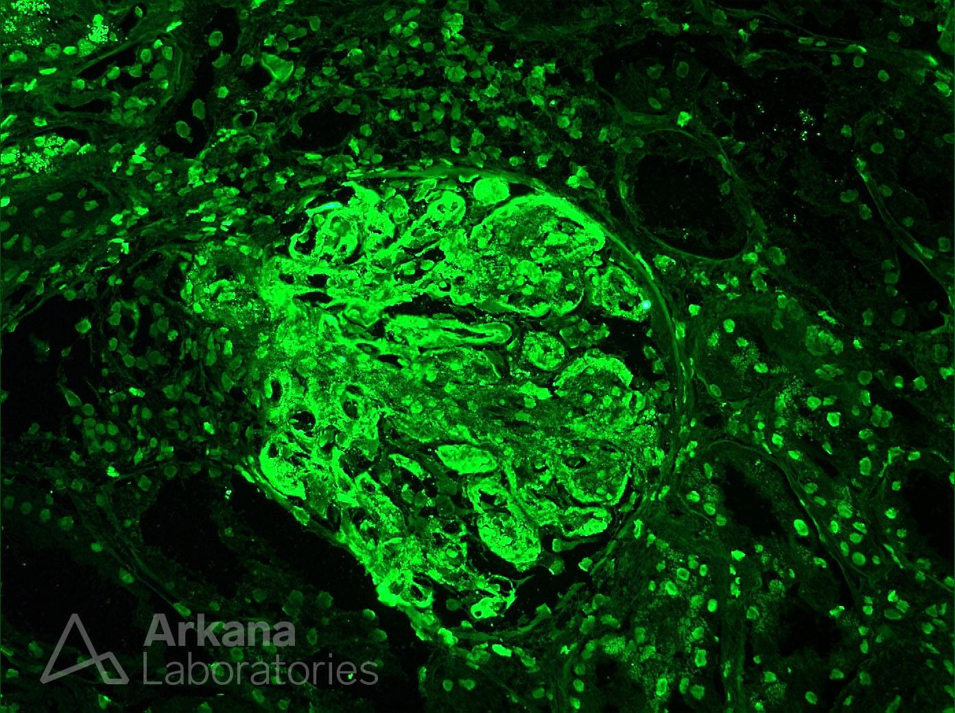
What is your diagnosis (IgG stain shown)?
The image show an IgG immunofluorescent stain with intense capillary wall and mesangial staining. While much of the capillary wall staining cannot be localized, some areas show a subendothelial pattern of deposition. Additionally, a tissue ANA pattern can be seen in the background. The presence of diffuse capillary wall and mesangial IgG staining with a tissue ANA pattern is highly suggestive of lupus nephritis which is the correct diagnosis in this case. Additionally, the presence of significant subendothelial located deposits is consistent with a proliferative lupus nephritis (ISN/RPS classes III and IV) which was correct in this case (diffuse lupus nephritis, ISN/RPS Class IV).
Quick note: This post is to be used for informational purposes only and does not constitute medical or health advice. Each person should consult their own doctor with respect to matters referenced. Arkana Laboratories assumes no liability for actions taken in reliance upon the information contained herein.