What is your diagnosis?
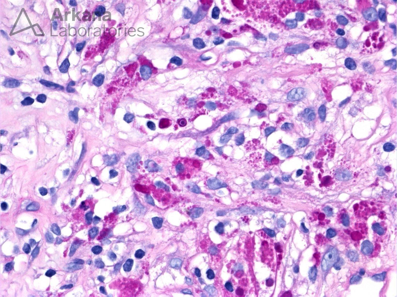
This photomicrograph shows a high power view of the renal interstitium with histiocytic inflammation and numerous histocytes with PAS-positive material within their cytoplasm. Within the center of the field is a small, target-shaped structure—a Michalis-Gutmann (MG) body. These findings are consistent with Malakoplakia which often forms mass lesions mimicking a neoplasm. Malakoplakia results from dysregulated lysosomal breakdown and processing of bacteria (typically E. coli) with resulting foamy macrophages with PAS-positive intracellular accumulations. Partially digested bacteria can also form a nidus for calcium and iron deposition, thus MG-body formation. Malakoplakia is an unusual finding and is not expected in an otherwise healthy person. It is frequently associated with an altered immune status such as HIV/AIDS, underlying malignancy, or immunosuppressive therapy.
Quick note: This post is to be used for informational purposes only and does not constitute medical or health advice. Each person should consult their own doctor with respect to matters referenced. Arkana Laboratories assumes no liability for actions taken in reliance upon the information contained herein.