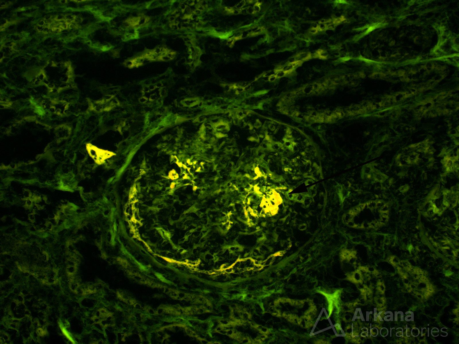
As published decades ago by Bonsib et al. (see reference), fluorescence microscopy of eosin-stained frozen section slides can be a valuable tool in providing diagnostic clues in kidney biopsies. The fluorescent properties of eosin dye allow for nice visualization of glomerular capillary and larger vascular basement membranes. Consequently, various disease processes which disrupt these structures can often be detected using eosin-stained frozen section slides. The presence of glomerular fibrin, for example, is easy to recognize using this method (see the image showing fibrinogen staining), and indicates a disease process causing glomerular capillary rupture, such as ANCA-related or immune complex-mediated glomerulonephritis. The extra time taken to examine such slides is usually well-spent and it just might uncover focal lesions not seen using routine light microscopy.
Bonsib SM, et al. Mod Pathol. 1990 Mar;3(2):204-10. Renal biopsy frozen section: a fluorescent study of hematoxylin and eosin-stained sections.
Quick note: This post is to be used for informational purposes only and does not constitute medical or health advice. Each person should consult their own doctor with respect to matters referenced. Arkana Laboratories assumes no liability for actions taken in reliance upon the information contained herein.