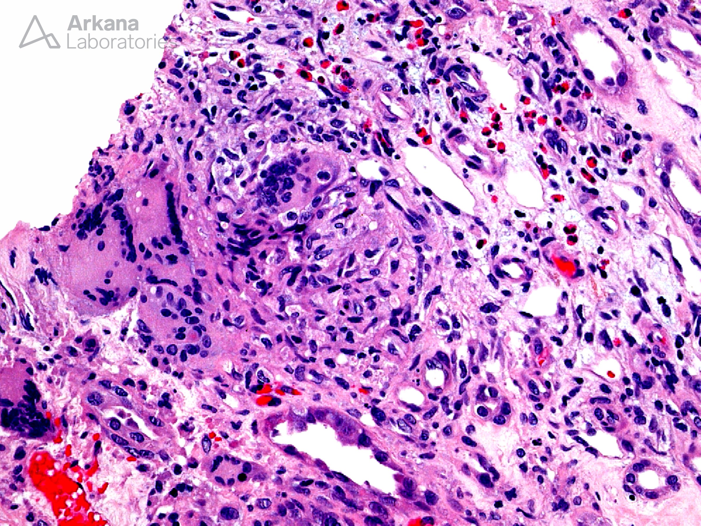
The photomicrograph shows an interstitial granuloma from a case of granulomatous interstitial nephritis. The presence of granulomas within interstitial nephritis frequently raises the differential diagnosis of unusual infections such as fungal and mycobacterial. However, a case series conducted within the United States showed that drug-induced reactions are the etiology of 45% of granulomatous interstitial nephritis cases followed by sarcoidosis in another 30%. The remaining 25% included 10% of cases that were idiopathic and 15% due to a variety of other etiologies such as systemic vasculitis, foreign body giant cell reaction, and infection.
Reference: Bijol V, et al. Int J Surg Pathol 14: 57-63, 2006
Quick note: This post is to be used for informational purposes only and does not constitute medical or health advice. Each person should consult their own doctor with respect to matters referenced. Arkana Laboratories assumes no liability for actions taken in reliance upon the information contained herein.