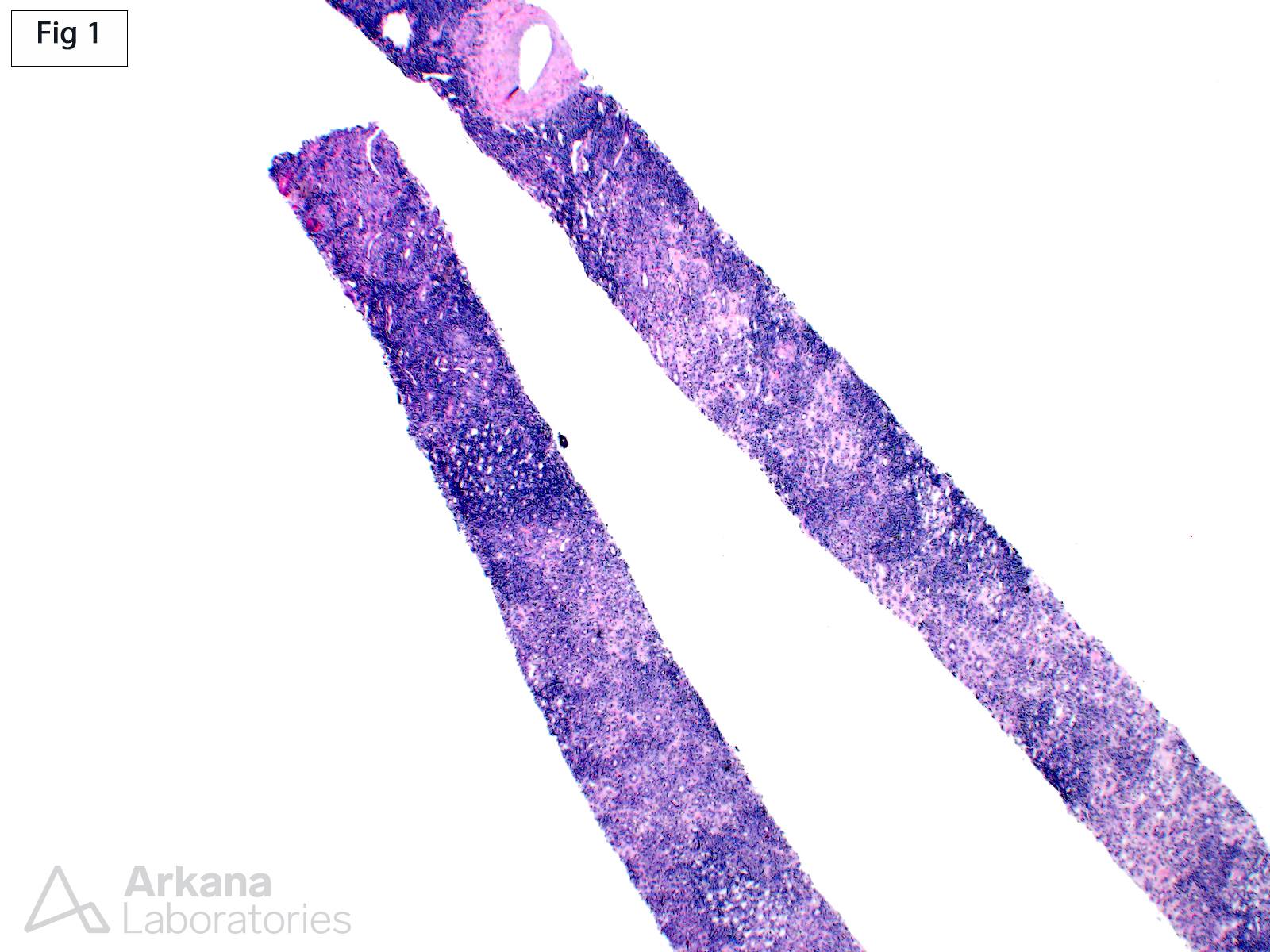
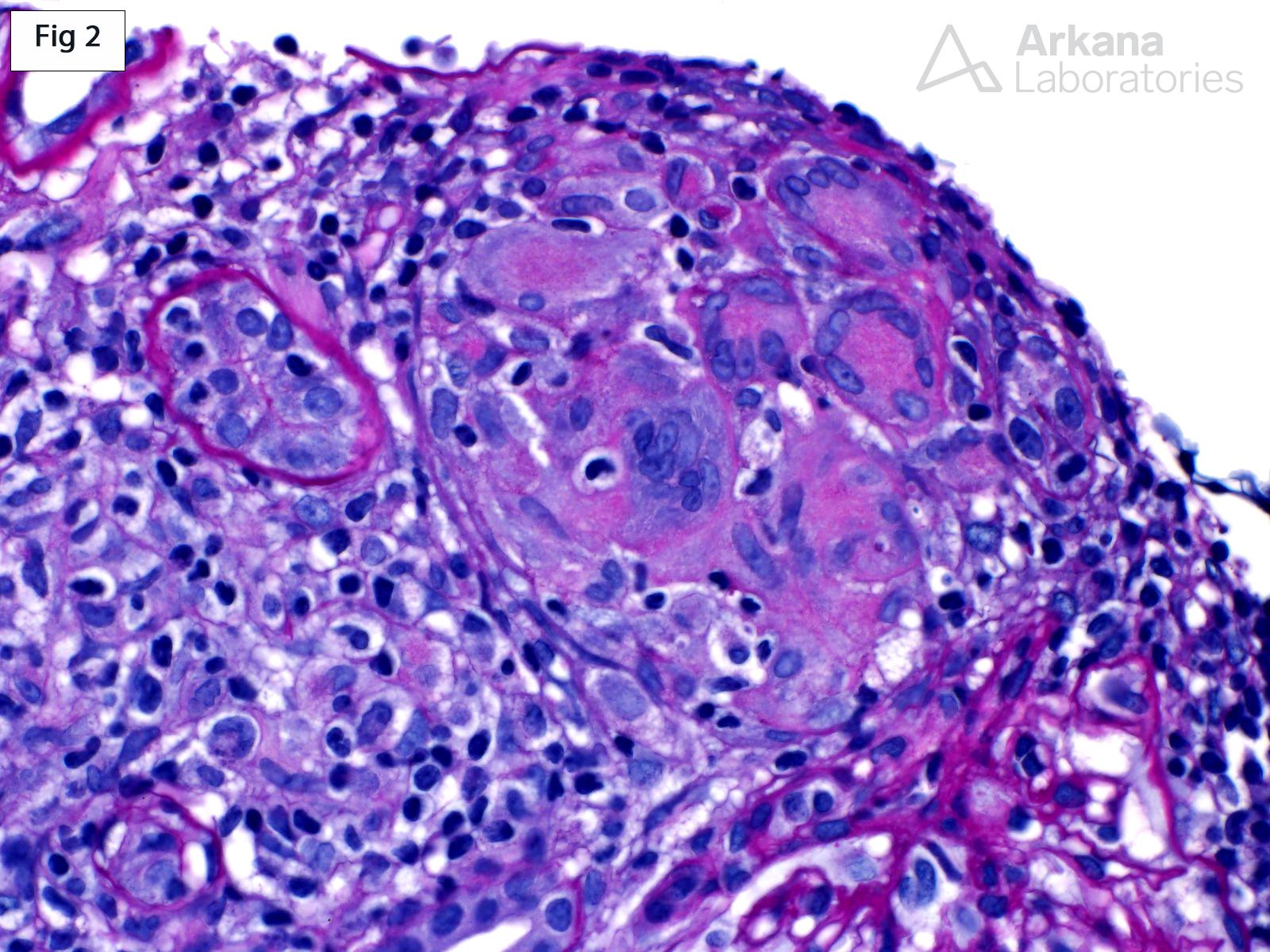
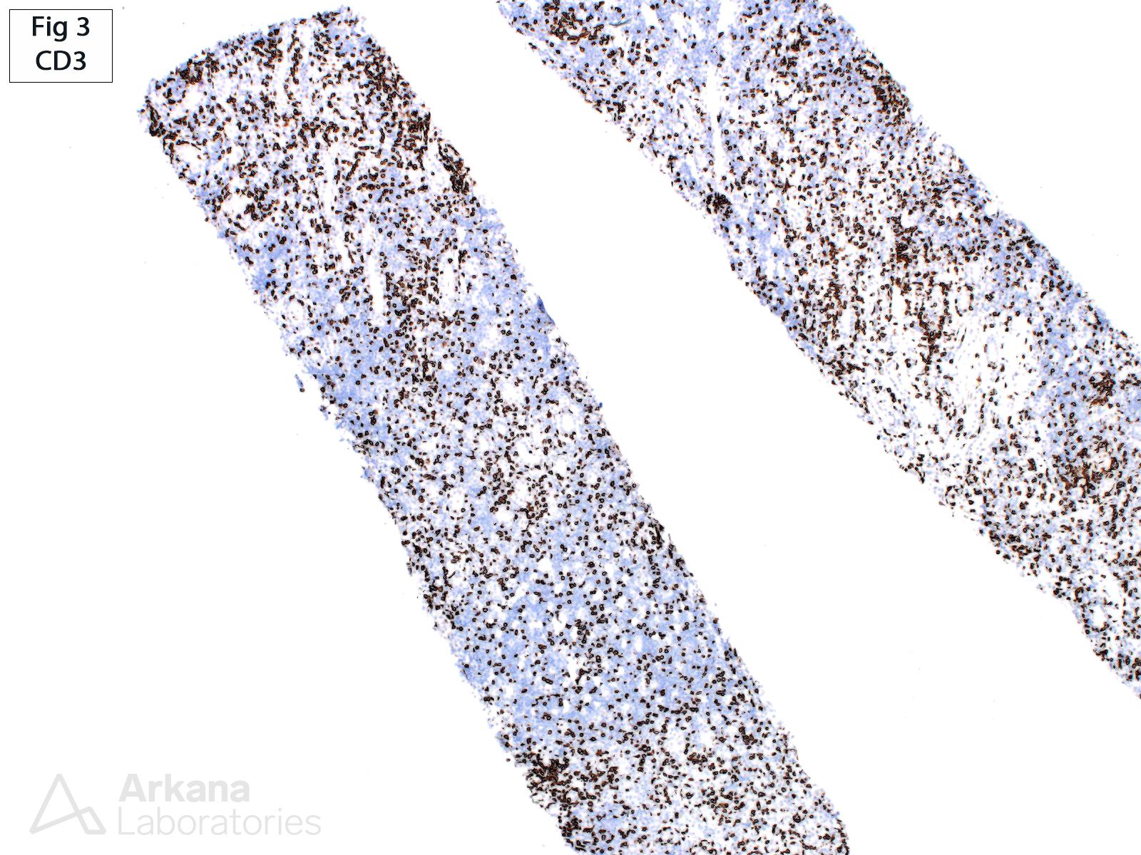
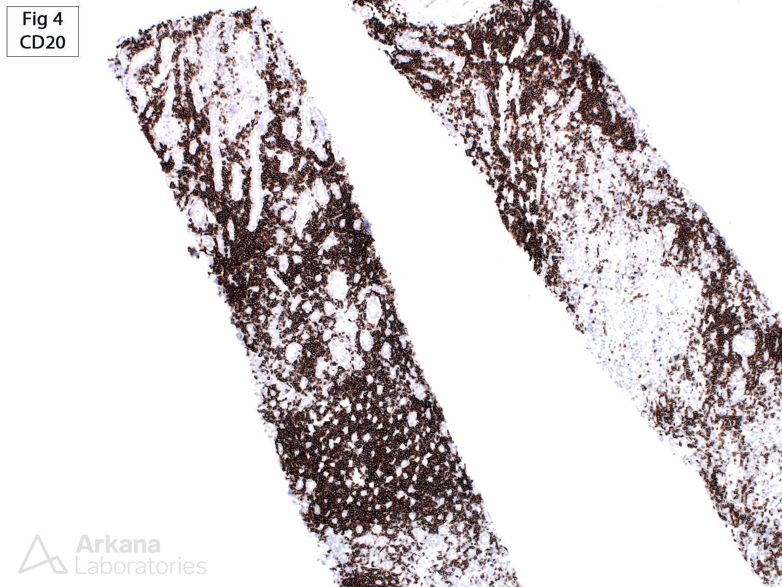
This is a renal biopsy from a 62-year-old female with a recent diagnosis of CLL/SLL, who presents with rapidly worsening renal function. The serum creatinine is 4.7 mg/dl (2.7 mg/dl 3 months prior). The biopsy shows a dense multifocal infiltrate of monomorphic small lymphocytes (Fig 1) which stain strongly positive for CD20 (Fig 4) and CD5 (not shown), and negative for CD3 (Fig 3). The background renal parenchyma shows moderate, mixed inflammation with scattered CD3 positive cells. Additionally, the interstitium multifocally shows granulomatous inflammation with giant cells (Fig 2). These findings are consistent with renal involvement by the patient’s know CLL/SLL along with a component of granulomatous interstitial nephritis. The differential diagnosis of granulomatous interstitial nephritis includes drug/hypersensitivity reaction, infections, autoimmune disorders, and sarcoidosis; however, it has also been reported in the setting of renal involvement by CLL/SLL (see reference).
Reference: Nasr SH, Shanafelt TD, Hanson CA, et al. Granulomatous interstitial nephritis secondary to chronic lymphocytic leukemia/small lymphocytic lymphoma. Ann Diagn Pathol. 2015;19:130-136.
Quick note: This post is to be used for informational purposes only and does not constitute medical or health advice. Each person should consult their own doctor with respect to matters referenced. Arkana Laboratories assumes no liability for actions taken in reliance upon the information contained herein.