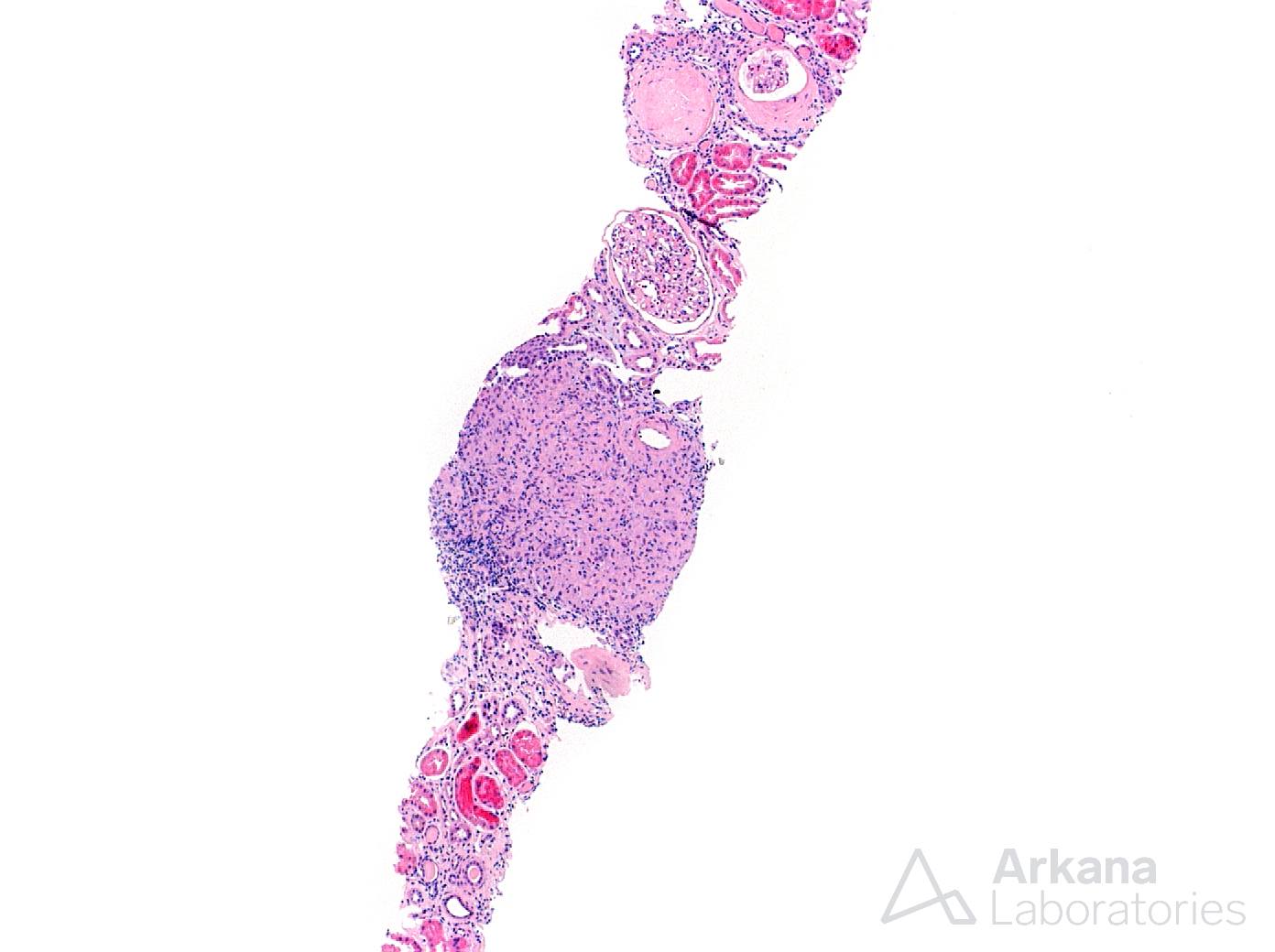
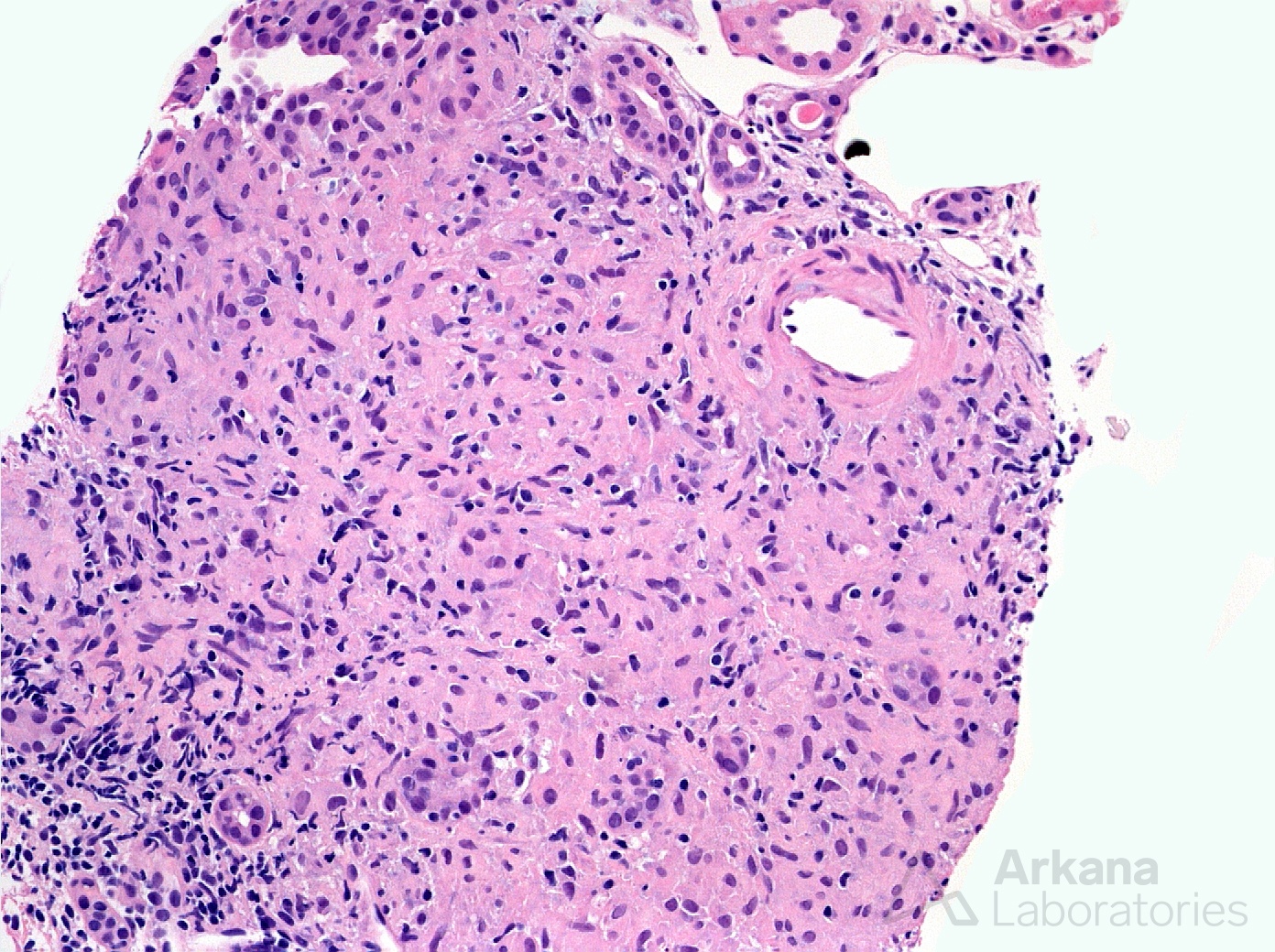
This biopsy is from a 25-year-old African American female with renal failure. The photomicrographs here show renal involvement by non-caseating granulomas eliciting the diagnosis of granulomatous interstitial nephritis. The patient was found to be hypercalcemic and to have hilar lymphadenopathy and reticulonodular infiltrates on chest x-ray and was diagnosed with sarcoidosis. A case series examining 46 cases of granulomatous interstitial nephritis (GIN) by Bijol et al (ref below) found the most common etiology (45%) of this pattern to be a drug-induced reaction. This was followed by sarcoidosis (29% of GIN), other (including infection) at 16% and there were 10% of cases that proved to be idiopathic. It should be pointed out that this case series was composed of cases from the United States and the etiologies from other parts of the world would likely be different.
Reference: Bijol V, et al. Int J Surg Pathol 14: 57-63, 2006
Quick note: This post is to be used for informational purposes only and does not constitute medical or health advice. Each person should consult their own doctor with respect to matters referenced. Arkana Laboratories assumes no liability for actions taken in reliance upon the information contained herein.