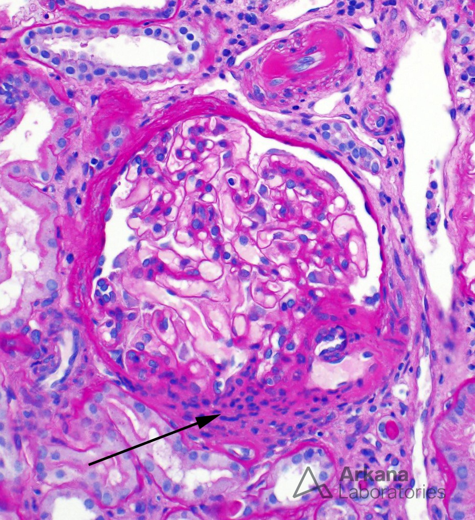
How do you identify the juxtaglomerular apparatus (JGA)?
The arrow in this image points to hyperplasia of the juxtaglomerular apparatus (JGA), which was seen diffusely in this biopsy. The JGA consists of vascular components (portions of the afferent and efferent arterioles), mesangial cell components (modified extraglomerular and intraglomerular smooth muscle cells), and a tubular component (the macula densa).
Diagnosis for juxtaglomerular apparatus hyperplasia
The differential diagnosis for JGA hyperplasia includes causes of chronic renal ischemia (e.g. renal artery stenosis or cardiac failure), various drugs (e.g. angiotensin II receptor antagonists and cyclosporine), and Bartter syndrome. This biopsy was taken from a child who had been diagnosed with Bartter syndrome approximately two years earlier – he originally presented with hypokalemia, alkalosis, hyperaldosteronism, and normal blood pressure. Since Bartter FC, et al. first described the syndrome in 1962 (https://www.ncbi.nlm.nih.gov/pubmed/13969763), at least six gene mutations affecting various tubular ion channels or transporters have been described.
Quick note: This post is to be used for informational purposes only and does not constitute medical or health advice. Each person should consult their own doctor with respect to matters referenced. Arkana Laboratories assumes no liability for actions taken in reliance upon the information contained herein.