Clinical History
This 77-year-old patient presented with atrophy of the cranial and extremity muscles, dysphagia, and multiple falls. Past medical history includes acute lymphoblastic lymphoma, marrow lymphoblasts 30%, and pancytopenia. Upon physical exam, cachectic appearance speech fluent without dysarthria, strength: 5-/5 upper extremities, 4+/5 lower extremities, MRI lumbar spine multilevel with degenerative changes and severe canal stenosis L3-4. EMG shows myopathic units of the abductor pulses bravos, pronator tarries, triceps and deltoid muscles. Nerve conduction study showed sensorimotor neuropathy. Lab results: aldolase 5.0, ANA negative, vitamin B12 normal, BNP elevated, C-reactive protein 2.0 (High), and serum protein electrophoresis showed a weak M-protein band thought clinically not to be clinically significant.
Question:
What is the pathological process?
A. Small artery vasculitis
B. Moyamoya disease
C. Obliterative vasculopathy
D. Vascular amyloidosis
Answer:
The correct answer is D. Vascular amyloidosis.
Bonus Question:
What is the underlying etiology?
A. Aβ deposition
B. Light chain disease
C. AA amyloidosis
D. Transthyretin deposition
Answer:
The correct answer is B. Light chain disease
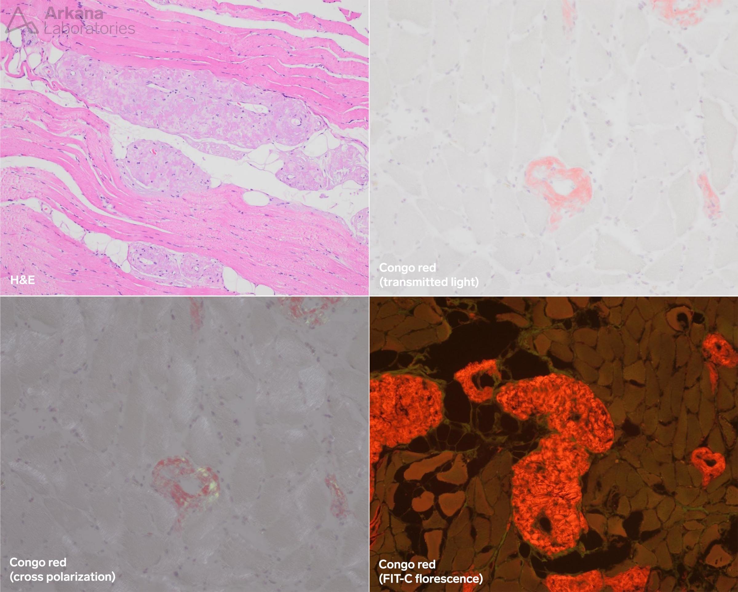
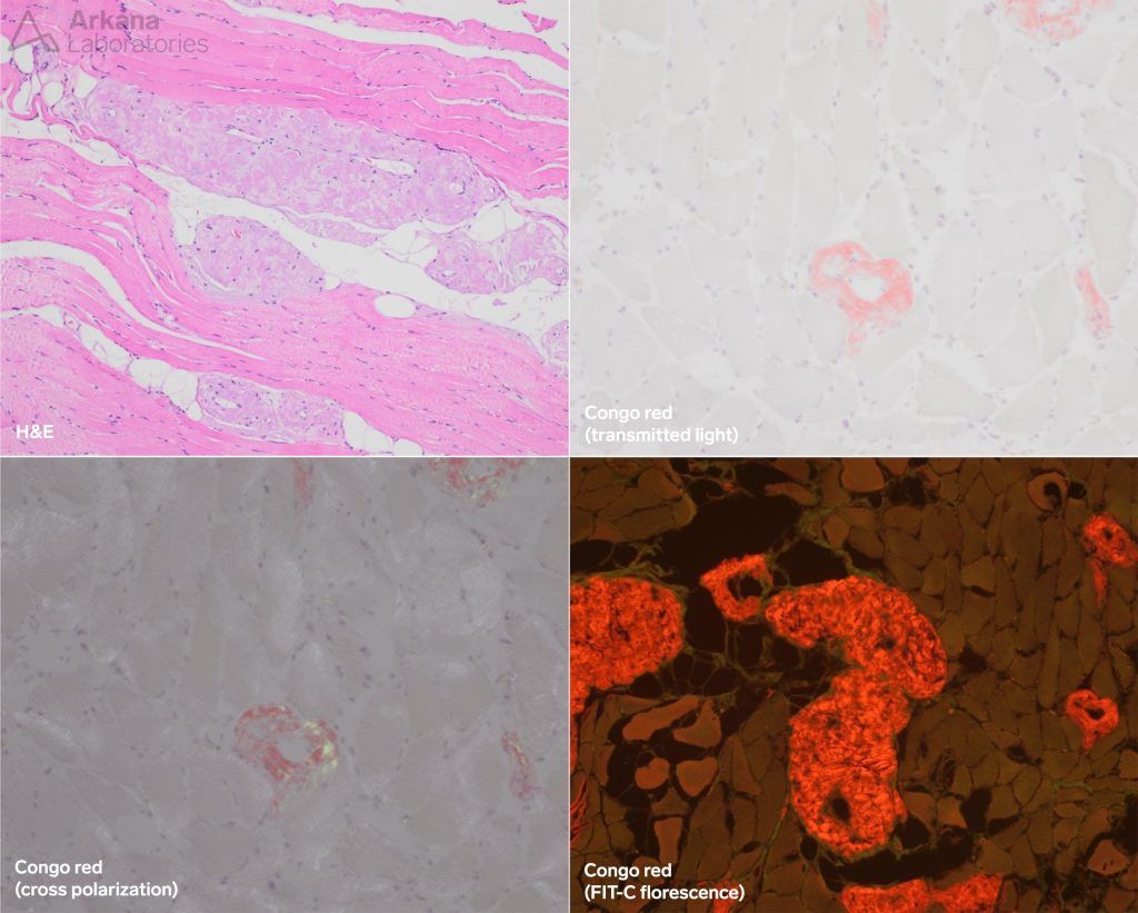
Vascular amyloidosis
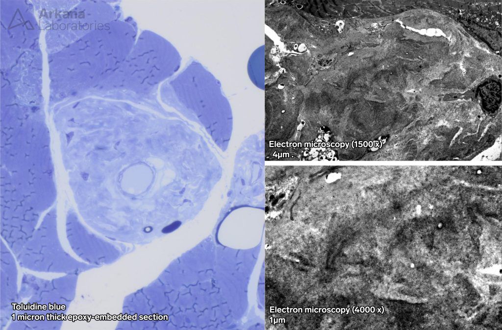
Amyloid- abnormal aggregation of amorphous protein of multiple types (immunoglobulins, Aβ, transthyretin, AA, etc.).
- Most are fibrillary as seen by electron microscopy, but a few, like the immunoglobulin- light chains kappa and lambda may not be.
- Can deposit almost anywhere but in muscle it is usually seen around vessels.
Congo Red- nonspecific stain to detect any amyloid (stains salmon or peach-orange)
- Shows characteristic “apple-green” birefringence under crossed-polarization
- Fluoresces red under Texas red or FIT-C fluorescence microscopy
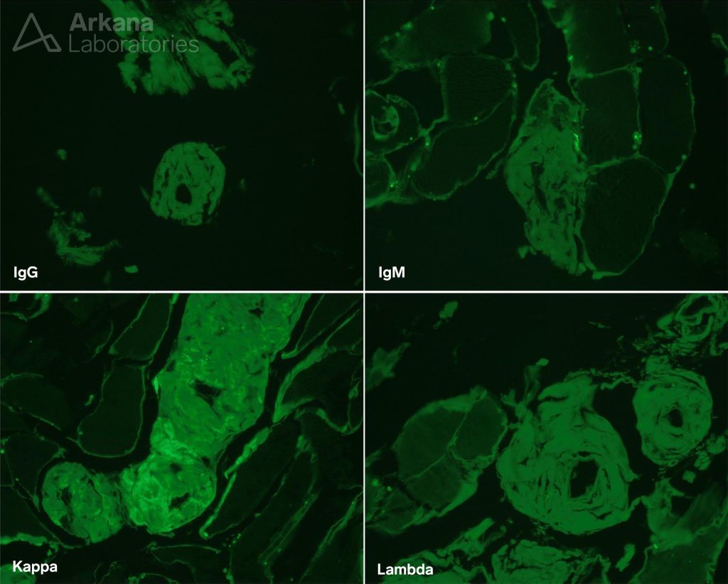
Typing amyloid
- Immuno-staining
- Here kappa restriction is demonstrated by immunofluorescence. This is most likely due to abnormal antibody shedding by the patient’s lymphoma
- Microdissection and mass spectroscopy can be used to confirm known proteins or identify unknown proteins
- Confirmed by mass spectroscopy
Quick note: This post is to be used for informational purposes only and does not constitute medical or health advice. Each person should consult their own doctor with respect to matters referenced. Arkana Laboratories assumes no liability for actions taken in reliance upon the information contained herein.