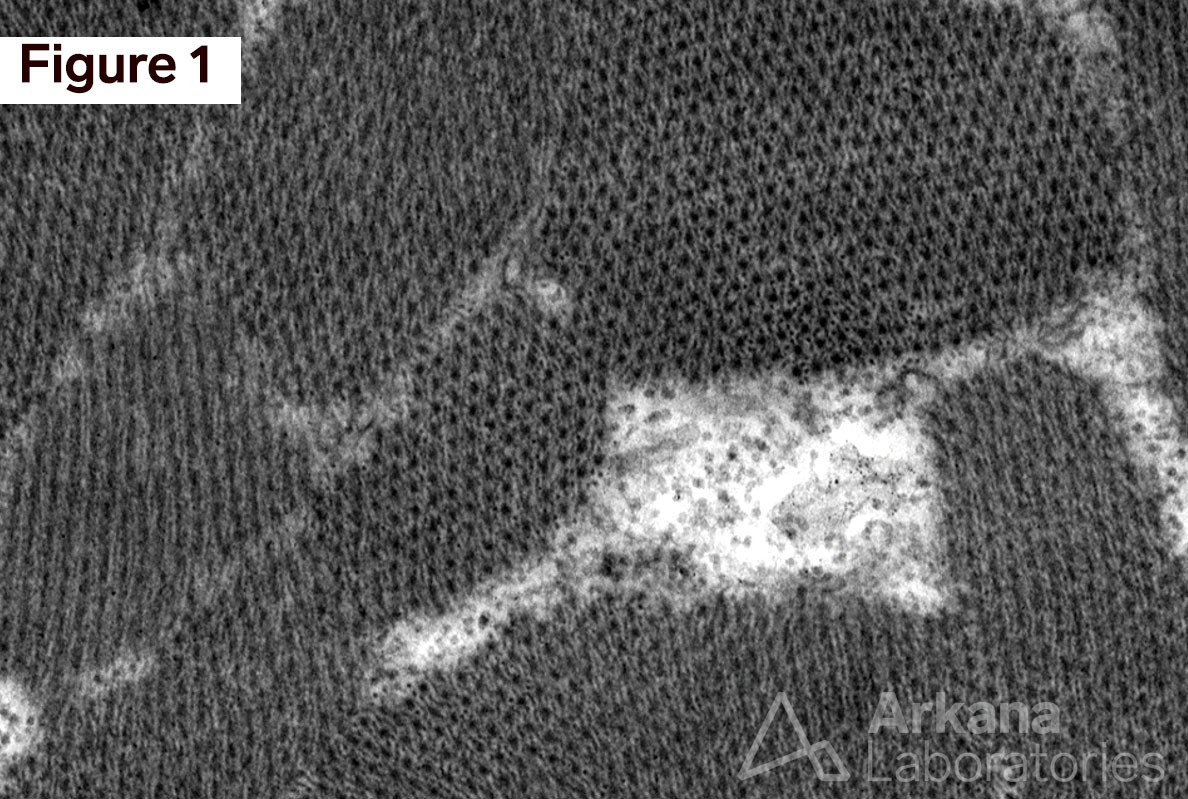
This week’s interesting image shows the examination of skeletal muscle in cross-section at the level of the A zone by electron microscopy in Figure 1, and corresponding illustration of the sarcomere in longitudinal section, Figure 2.
Figure 1. Ultrastructure of sarcomere bundles. The contractile elements of skeletal muscle are seen in cross-section at the level where myosin thick filaments and actin thin filaments overlap at the “anisotropic” or “A” zone of the sarcomere. Electron Microscopy (EM): 25000x original magnification.
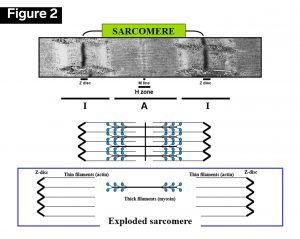
Figure 2. Illustration of sarcomere bundles. The sarcomere contractile apparatus viewed in longitudinal section shows various degrees of overlap of the thick (myosin) and thin filaments (actin) in the A zone depending on the degree of muscle contraction. Illustration courtesy of Dr. Jon Wilson.
Quick note: This post is to be used for informational purposes only and does not constitute medical or health advice. Each person should consult their own doctor with respect to matters referenced. Arkana Laboratories assumes no liability for actions taken in reliance upon the information contained herein.