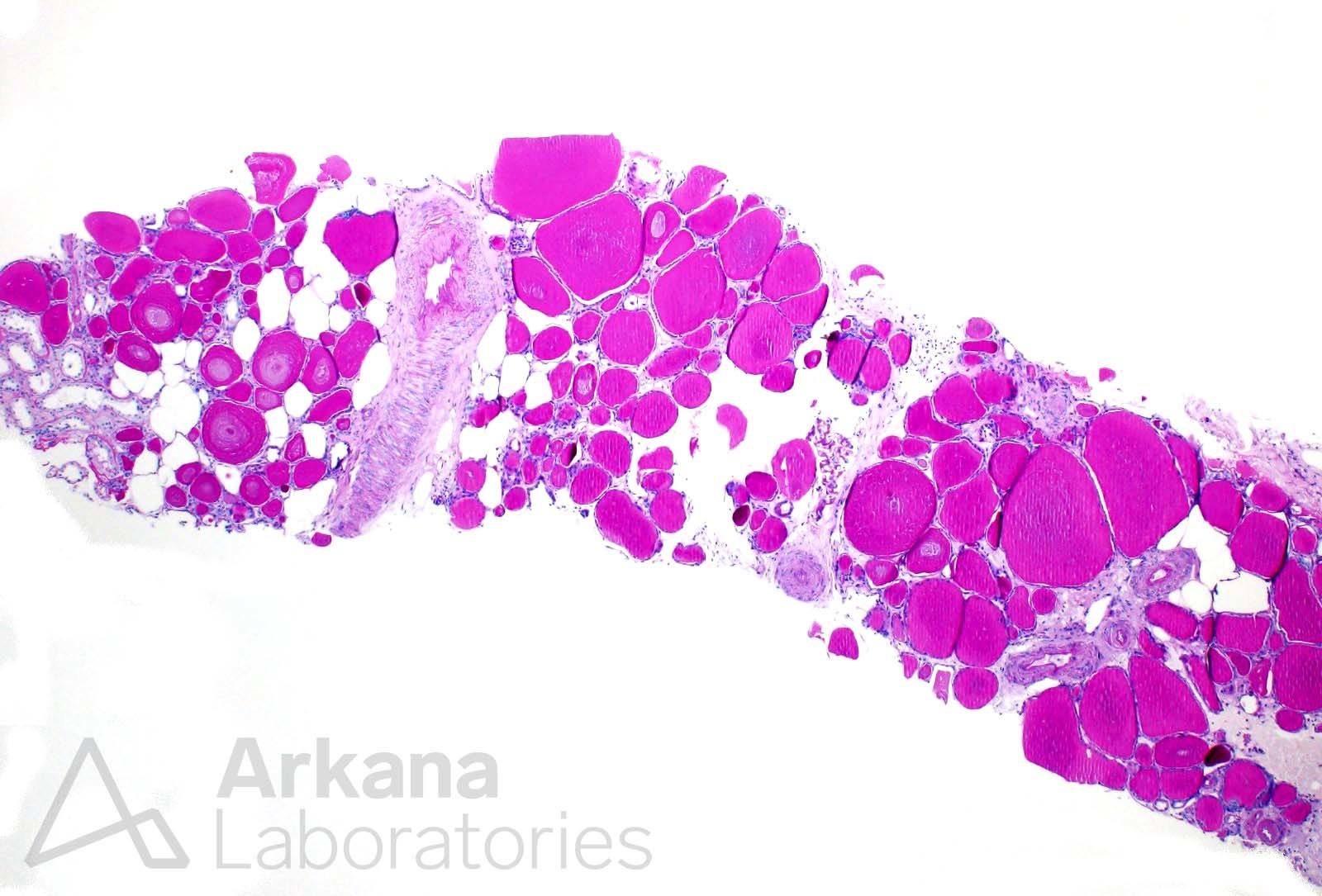
One of the many patterns of tubular atrophy in the kidney is the aptly named “thyroidization” pattern because of its resemblance to normal thyroid gland follicles. The dilated tubules contain abundant protein, resembling thyroid colloid, which is surrounded by flattened epithelial cells. This pattern of tubular atrophy is non-specific, although it is often more frequently encountered in the setting of chronic pyelonephritis and reflux nephropathy.
Reference
Lusco MA, Fogo AB, Najafian B, Alpers CE. AJKD Atlas of Renal Pathology: Tubular Atrophy. Am J Kidney Dis. 2016 Jun; 67(6):e33-4.
Quick note: This post is to be used for informational purposes only and does not constitute medical or health advice. Each person should consult their own doctor with respect to matters referenced. Arkana Laboratories assumes no liability for actions taken in reliance upon the information contained herein.