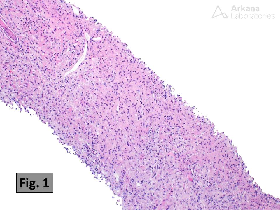
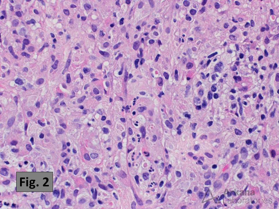
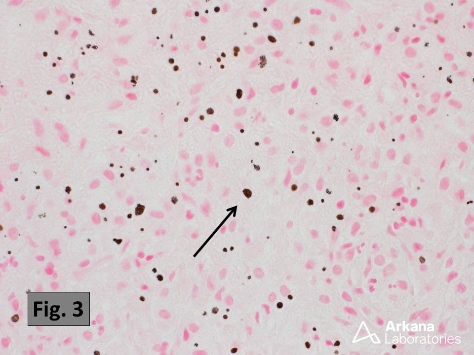
This kidney biopsy shows the histologic features characteristic of malakoplakia, which includes a dense, often obliterative, inflammatory infiltrate rich in histiocytes (Fig. 1 and 2) with numerous small intracytoplasmic concretions known as Michaelis-Gutmann bodies (these are best identified using histochemical staining for calcium as in Fig. 3). Malakoplakia is thought to result from chronic bacterial infection of the urinary tract (the most common causative organism is E. coli). Many cases are associated with the formation of small plaques and/or mass lesions. The bladder is the most common site of involvement, but kidney involvement occurs in about 15% of cases. The differential diagnosis includes xanthogranulomatous pyelonephritis.
Reference:
Kobayashi A, et al. Malakoplakia of the kidney. Am J Kid Dis. 2008 Feb;51(2):326-30. PMID: 18215711.
Quick note: This post is to be used for informational purposes only and does not constitute medical or health advice. Each person should consult their own doctor with respect to matters referenced. Arkana Laboratories assumes no liability for actions taken in reliance upon the information contained herein.