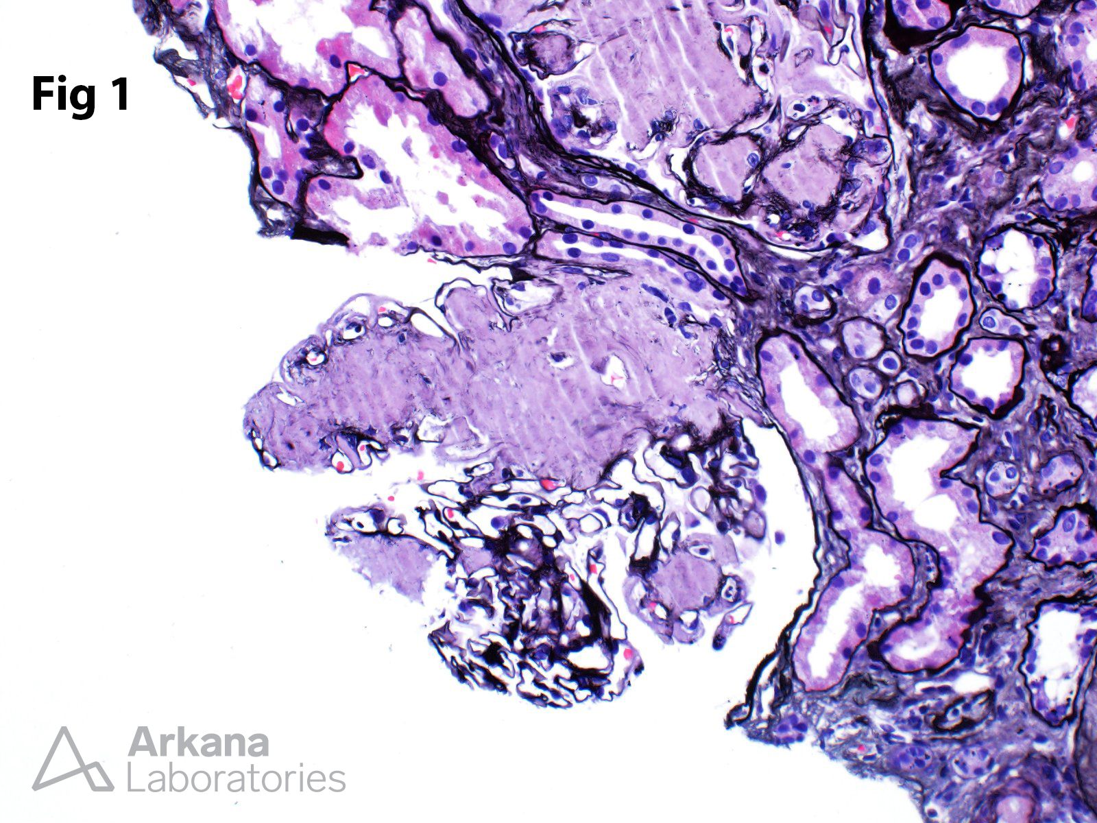
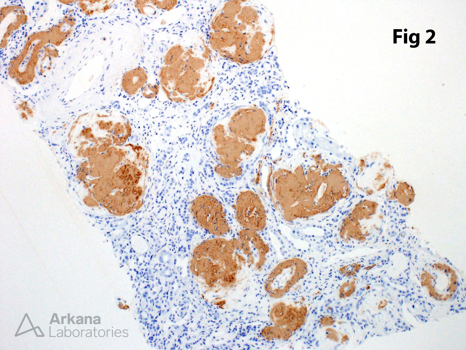
This biopsy from a patient with massive proteinuria showed diffuse and near-global involvement of glomeruli by amorphous deposits (Fig. 1) which were Congo red positive (not shown). The amyloid subtype was confirmed as AA amyloid using immunohistochemistry (Fig. 2). Immunofluorescence studies showed no evidence of kappa or lambda light chain restriction within the deposits (not shown). AA amyloidosis is usually diagnosed in the setting of a prolonged inflammatory condition along with high levels of serum amyloid A (SAA) precursor protein, from which the AA amyloid fibril protein is derived. Since the early twentieth century, the incidence of AA amyloidosis has decreased with the emergence of more effective treatments for chronic infectious and autoimmune diseases.
Reference: Westermark GT, et al. Annual Rev Pathol. 2015; 10:321-44.
Quick note: This post is to be used for informational purposes only and does not constitute medical or health advice. Each person should consult their own doctor with respect to matters referenced. Arkana Laboratories assumes no liability for actions taken in reliance upon the information contained herein.