Search & Filter All Posts
Results for Neuro Notes
(140 Results)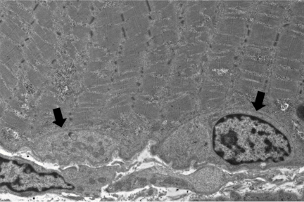
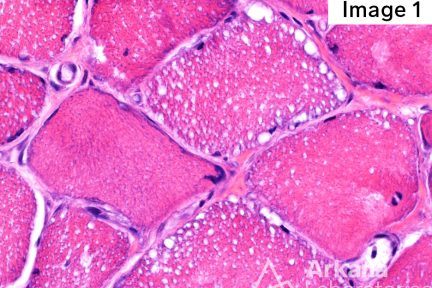
Satellite Cells
An adult patient with proximal muscle weakness presents with CK 4400 and skeletal muscle biopsy of the thigh shows frequent…
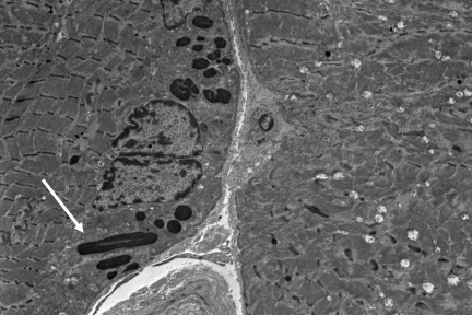
Nemaline Rods
What are these dark (osmophilic) structures seen in this electron micrograph of skeletal muscle? These osmophilic structures are Nemaline…
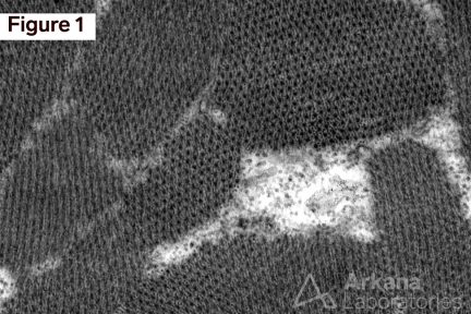
Sarcomere Bundles of the A Zone
This weeks interesting image shows the examination of skeletal muscle in cross-section at the level of the A zone by…
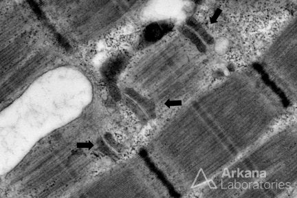
Tangential Sections Through Three Triads
Can you identify the structure(s) identified by arrows in this muscle biopsy? Tangential sections through three triads (pale T-tubules…
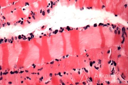
Contraction Band Artifact
Contraction band artifact (Hematoxylin and Eosin stained section from formalin fixed paraffin embedded tissue 400x magnification) This skeletal muscle…
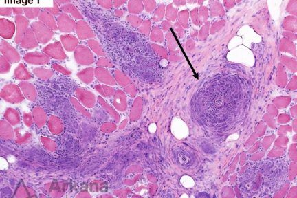
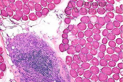
Sarcoid Myopathy and Vasculopathy
What is your diagnosis and how would you characterize the designated lesions in Figures 1 & 2? (Black Arrows) Hematoxylin…
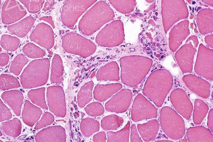
Grouped Atrophy
This skeletal muscle biopsy shows grouped atrophy, which is a finding characteristic of denervation. This morphologic alteration indicates the presence…
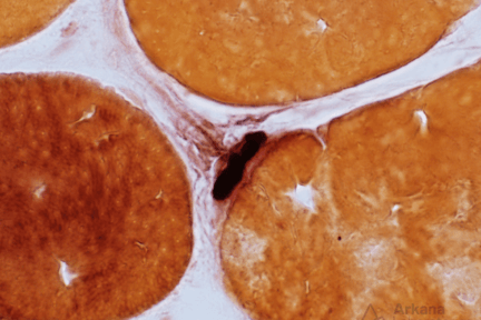
The Neuromuscular Junction
What is the dark staining structure? (Hint: note the small nerve twig associated with the structure). The neuromuscular junction (NMJ;…
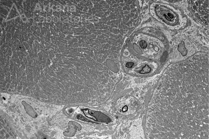
Nerve Twigs Within Endomysial Connective Tissue
This week’s Neuro Notes shows ultrastructural examination of a muscle biopsy can sometimes yield additional useful information, such as evaluation…
Macrophagic Myofasciitis (MMF)
This previously healthy pediatric patient presented with peculiar facial muscle hypertrophy, and genetic testing revealed one pathologic variant and two…
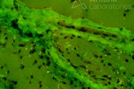
Other Uses For the Congo Red Stain
Did you know there could be other uses for Congo Red Stain? Additional unintended useful information can be obtained from…
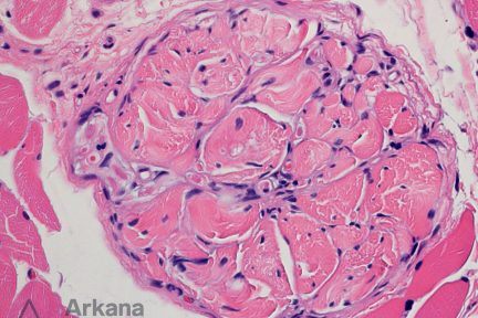
Golgi Tendon Organ
In reviewing this skeletal muscle biopsy, what is this structure and what is its function? (Hematoxylin and Eosin stained FFPE…
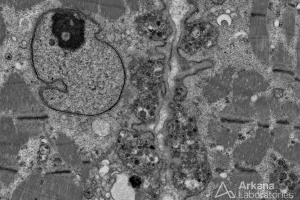
Exocytosis in a Skeletal Muscle Biopsy
This electron micrograph of two adjacent skeletal muscle cells demonstrates simultaneous exocytosis. Exocytosis in this case shows extravasation of lysosomes…
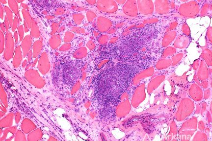
Sarcoidosis in a Skeletal Muscle Biopsy
These images show skeletal muscle from the thigh of an African-American patient with muscle weakness and elevated CPK levels. Occasional…
Lipid Storage Myopathy
This adult patient presented with progressive muscle weakness, elevated CK, and was admitted for rhabdomyolysis. Skeletal muscle biopsy was performed…