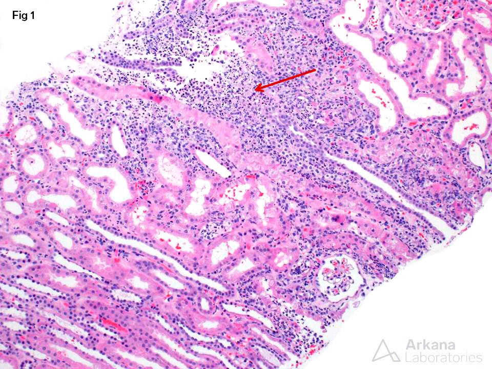
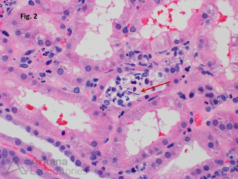
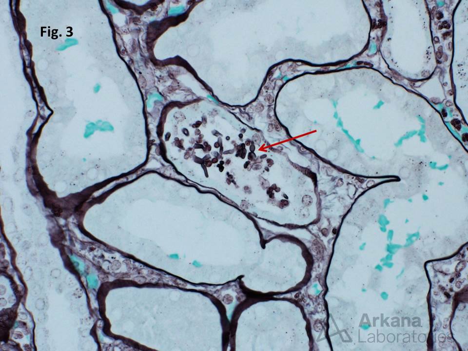
Cortical microabscess formation is a clue to the presence of an infectious organism. Various bacteria, viruses, fungi, and even parasites may cause similar appearing lesions. This biopsy is taken from a renal transplant patient with acute kidney injury and microscopic hematuria. The low-power image shows a densely mixed cellular infiltrate involving the tubulointerstitium (Fig. 1). Higher magnification shows tubulitis with intratubular leukocytes and rare structures suspicious for fungal elements (Fig. 2). Gomori methenamine silver staining confirms the presence of budding yeast forms (Fig. 3 arrow) with hyphae and pseudohyphae. Fungal isolates from urine culture showed colony morphology characteristics consistent with Candida species.
Quick note: This post is to be used for informational purposes only and does not constitute medical or health advice. Each person should consult their own doctor with respect to matters referenced. Arkana Laboratories assumes no liability for actions taken in reliance upon the information contained herein.