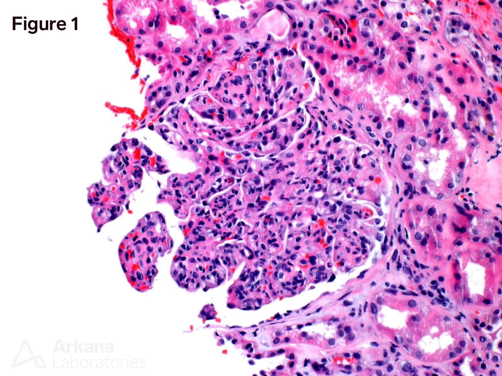
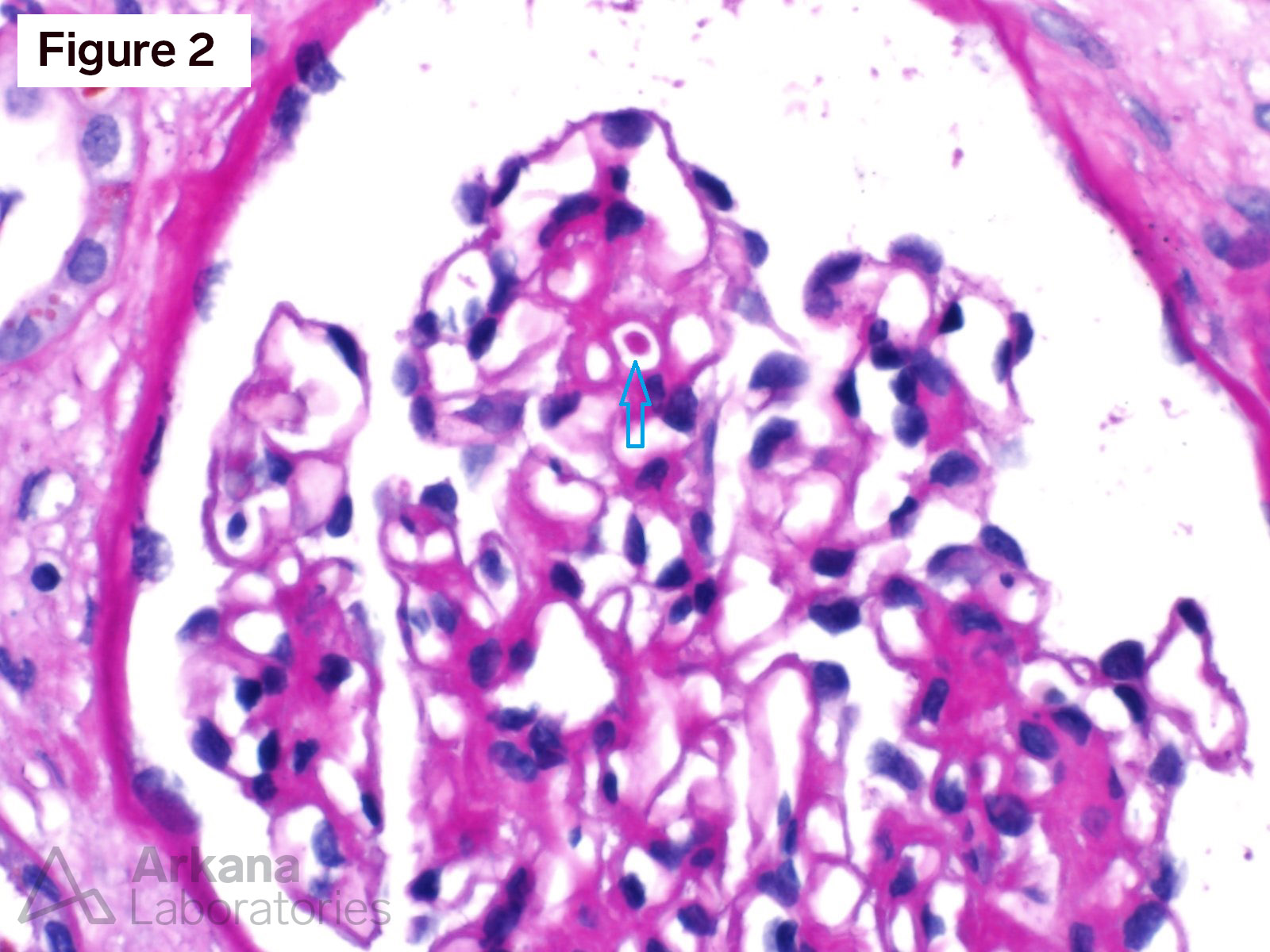
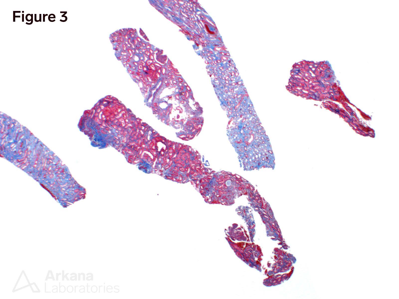
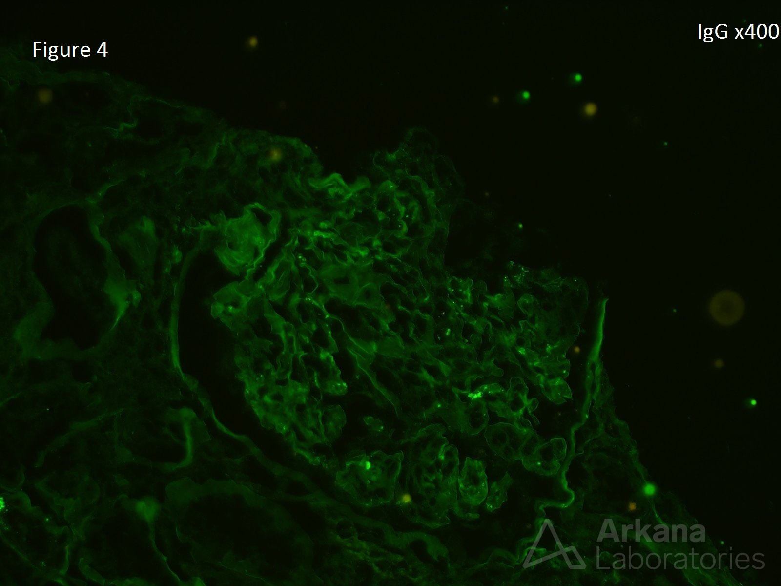
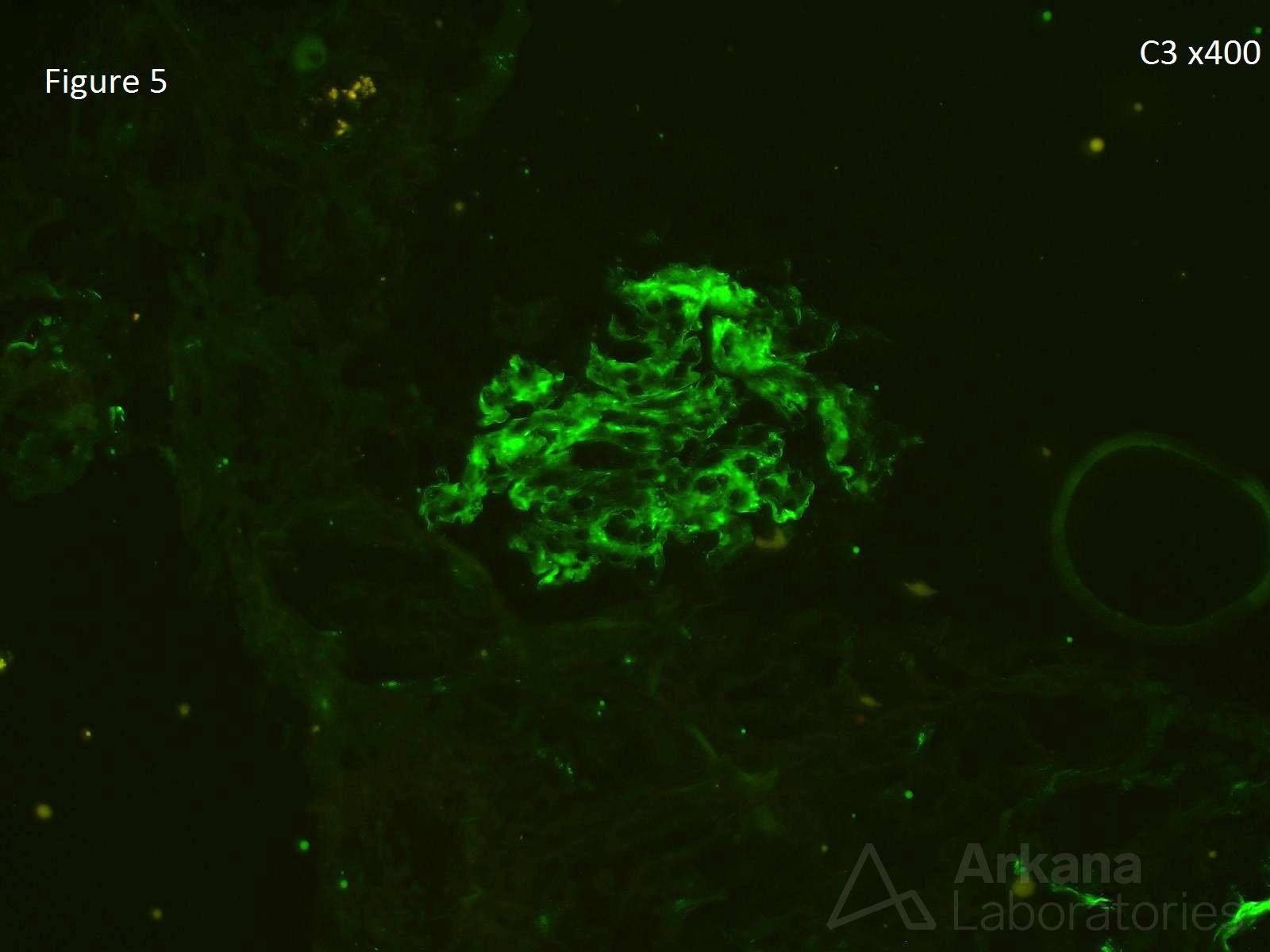
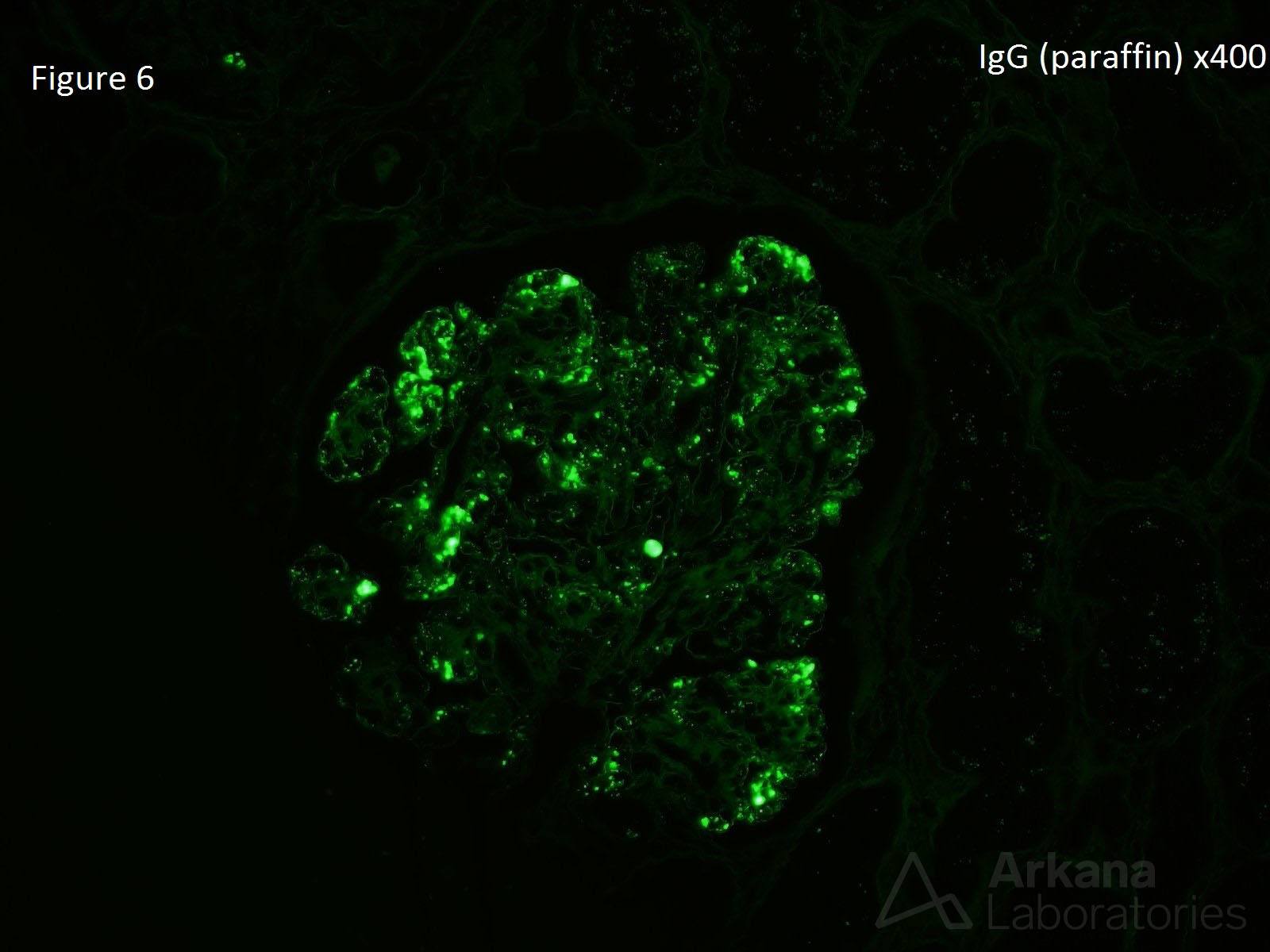
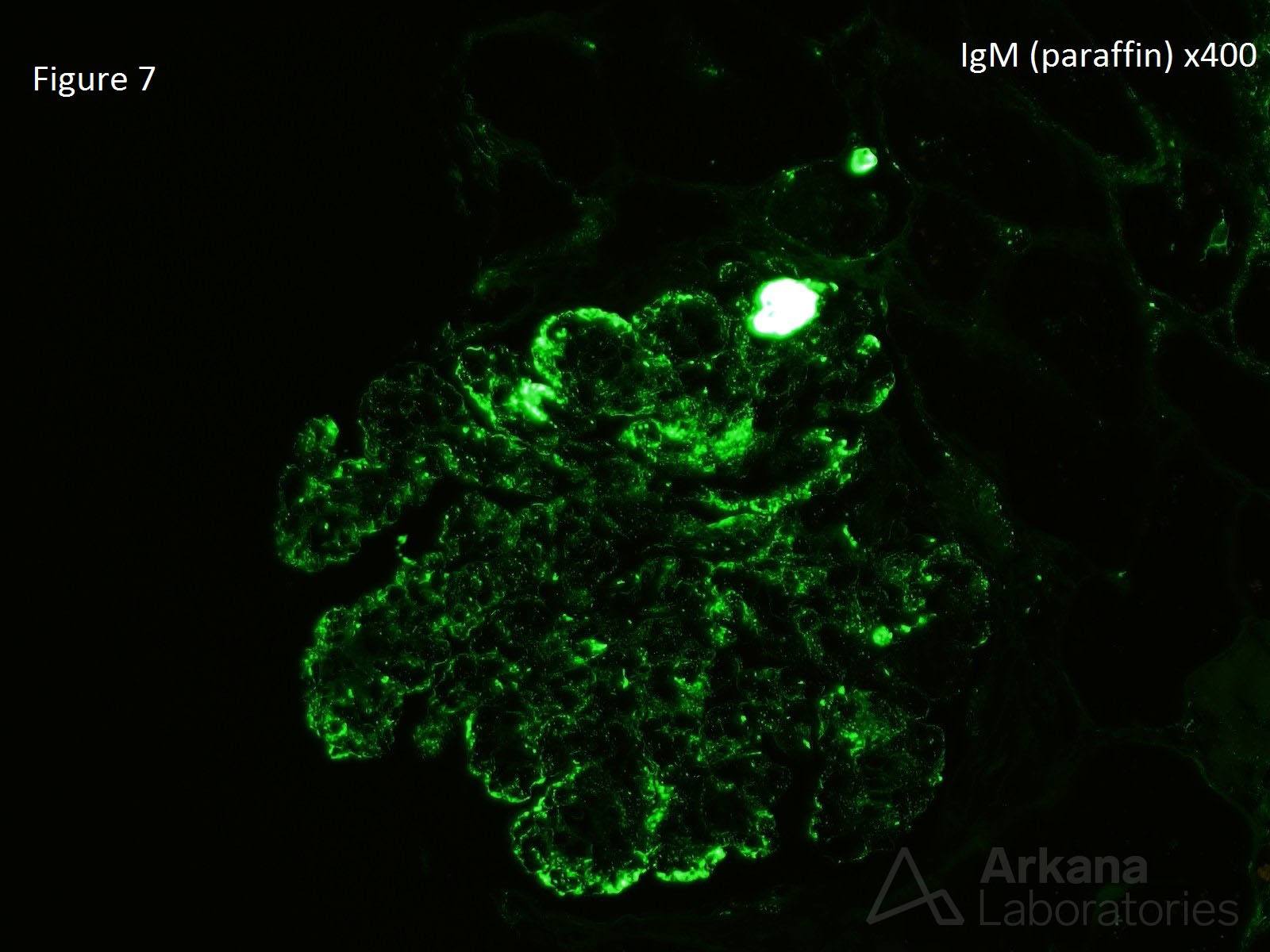
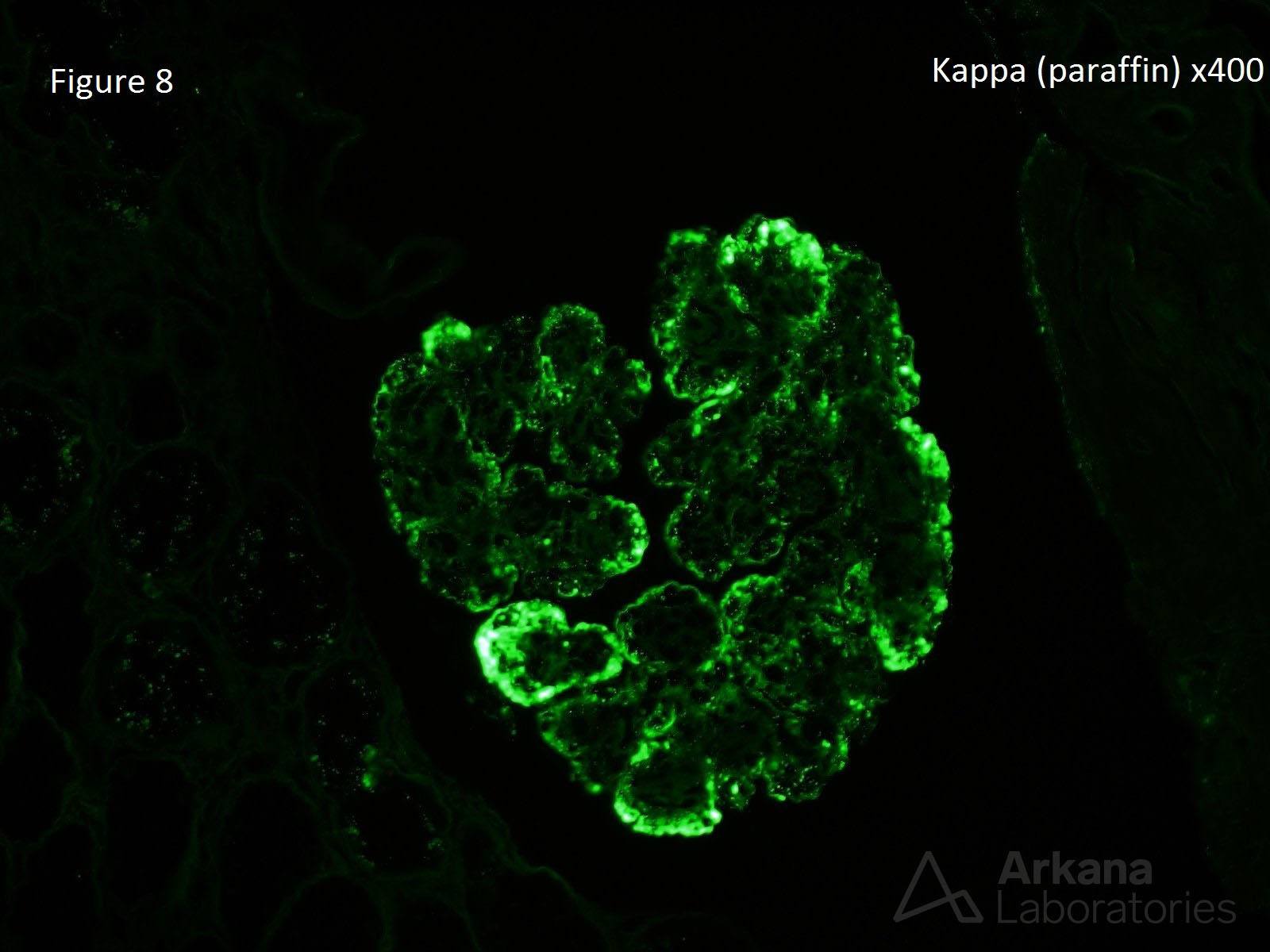
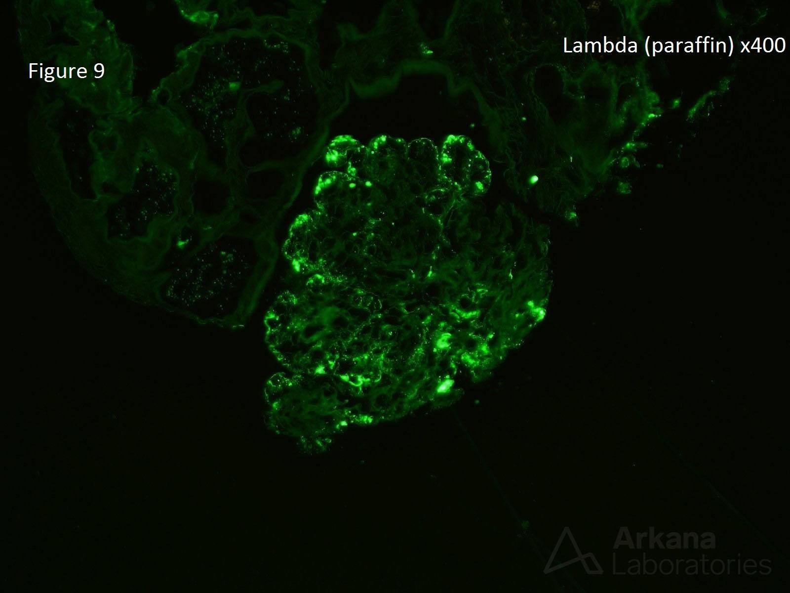
The patient is a 60-year-old male who presents with proteinuria, hematuria, and a creatinine of 2.8 mg/dL. He was in his normal state of health until 2 weeks ago when he noticed he was having trouble putting on his shoes. Serologies show low complements and an albumin of 2.3 g/dL. Serologies for ANA, c-ANCA, p-ANCA, hepatitis B, and hepatitis C are negative. No monoclonal spike is found on serum electrophoresis. Figure 1 shows accentuation of lobulation with both mesangial and endocapillary hypercellularity. This is a membranoproliferative pattern. Figure 2 shows a small, hyaline thrombus. Figure 3 shows moderate interstitial fibrosis. Figure 4 shows no staining for IgG (also of note: IgA, IgM, kappa and lambda light chains are negative). Figure 5 shows staining for C3. Figures 6, 7, 8, & 9 shows immunofluorescence staining on retrieved, pronase digested, paraffin-embedded tissue for IgG, IgM, kappa, and lambda light chains, respectively. This is a case of noninfectious, mixed cryoglobulinemic glomerulonephritis with renal involvement.
Most of us think of cryoglobulinemic glomerulonephritis solely related to hepatitis B or hepatitis C. However, there is a subset of cryoglobulinemic glomerulonephritis where an underlying infectious etiology cannot be identified. Approximately 22% of the noninfectious renal mixed cryoglobulinemic glomerulonephritis are related to Sjögren’s, 28% related to a lymphoproliferative disorder, and 50% are considered essential. Approximately 90% of cases of noninfectious renal mixed cryoglobulinemic glomerulonephritis have a MPGN pattern as illustrated in figure 1. Intraluminal thrombi are observed in approximately half of biopsies as shown in figure 2. The only immunofluorescence stain on frozen tissue to show positivity in this case was C3 (figure 5). “Un-masking” of heavy and light chains can be seen when immunofluorescence is performed on the paraffin-embedded tissue (figure 4, figure 6, figure 7, figure 8, figure 9).
https://www.ncbi.nlm.nih.gov/pubmed/26260165
Quick note: This post is to be used for informational purposes only and does not constitute medical or health advice. Each person should consult their own doctor with respect to matters referenced. Arkana Laboratories assumes no liability for actions taken in reliance upon the information contained herein.