Clinical History:
- This 40-year-old patient presented with one month history of progressive muscle weakness, joint pain, and skin rash
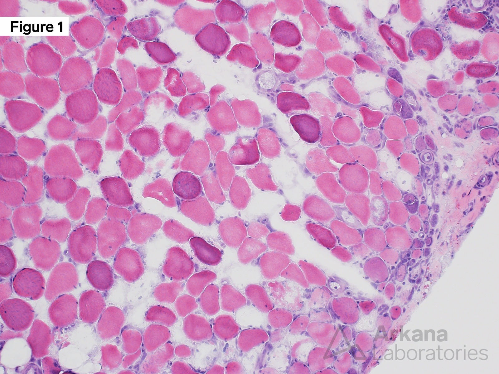
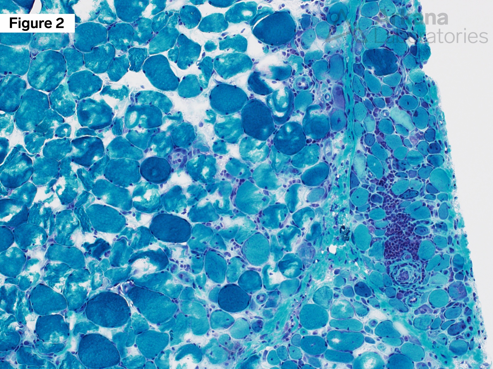
- MRI showed prominent muscle edema throughout the right and left thighs
- Physical examination demonstrated a skin rash involving their torso, face, and neck
- Laboratory studies showed elevated CPK (45o), CRP (1.09) and sedimentation rate (79), positive ANA and dsDNA, and negative Jo-1 autoantibody
- Myositis specific antibody panel test results were pending. They were treated with prednisone prior to muscle biopsy and reported some improvement in their symptoms
Question:
Positive immunohistochemical staining for MxA (myxovirus resistance protein A) is most commonly seen in the setting of which of the following?
A. Dermatomyositis
B. Polymyositis
C. Antisynthetase syndrome
D. Inclusion Body Myositis

Answer:
Positive immunohistochemical staining for MxA (myxovirus resistance protein A) is most commonly seen in the setting of which of the following?
A. Dermatomyositis <– correct answer
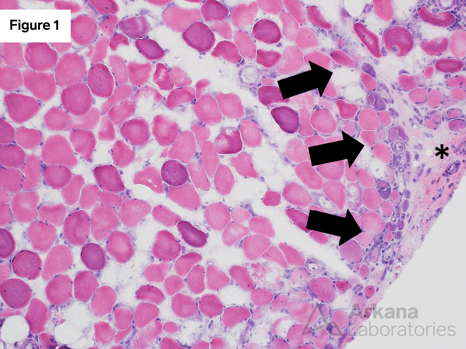
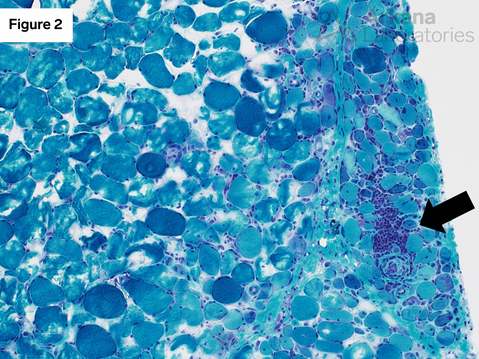
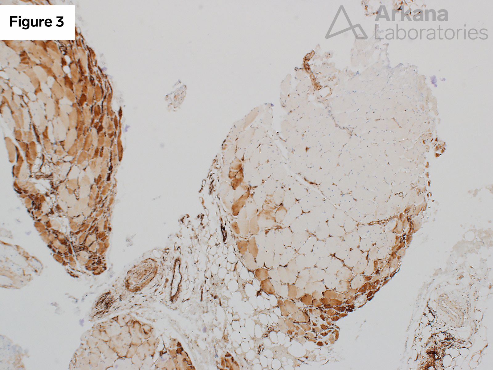
MxA (myxovirus resistance protein A) is a type I interferon (IFN) inducible gene product, with increased expression in the setting of viral infection. Increased expression is also seen in the setting of dermatomycosis and serves as a useful diagnostic aid. The inflammatory myopathy related to anti-synthetase syndrome may also show a perifascicular pattern of muscle fiber injury (perifascicular atrophy), but does not typically show increased expression of MxA.
Reference(s) / additional reading:
- Bolko L, Jiang W, Tawara N, Landon-Cardinal O, AnquetilC, Benveniste O, Allenbach Y. The role of interferons type I, II and III in myositis: A review. Brain Pathol. 2021 May;31(3):e12955. doi: 10.1111/bpa.12955. PMID: 34043262; PMCID: PMC8412069.
- Uruha A, Allenbach Y, Charuel JL, Musset L, Aussy A, Boyer O, Mariampillai K, Landon-Cardinal O, Rasmussen C, Bolko L, Maisonobe T, Leonard-Louis S, Suzuki S, Nishino I, Stenzel W, Benveniste O. Diagnostic potential of sarcoplasmic myxovirus resistance protein A expression in subsets of dermatomyositis. Neuropathol ApplNeurobiol. 2019 Aug;45(5):513-522. doi: 10.1111/nan.12519. Epub 2018 Nov 22. PMID: 30267437.
-
Tanboon J, Inoue M, Saito Y, Tachimori H, Hayashi S, Noguchi S, Okiyama N, Fujimoto M, Nishino I. Dermatomyositis: Muscle Pathology According to Antibody Subtypes. Neurology. 2022 Feb 15;98(7):e739-e749. doi: 10.1212/WNL.0000000000013176. Epub 2021 Dec 6. PMID: 34873015; PMCID: PMC8865893.
Quick note: This post is to be used for informational purposes only and does not constitute medical or health advice. Each person should consult their own doctor with respect to matters referenced. Arkana Laboratories assumes no liability for actions taken in reliance upon the information contained herein.