What is your diagnosis?
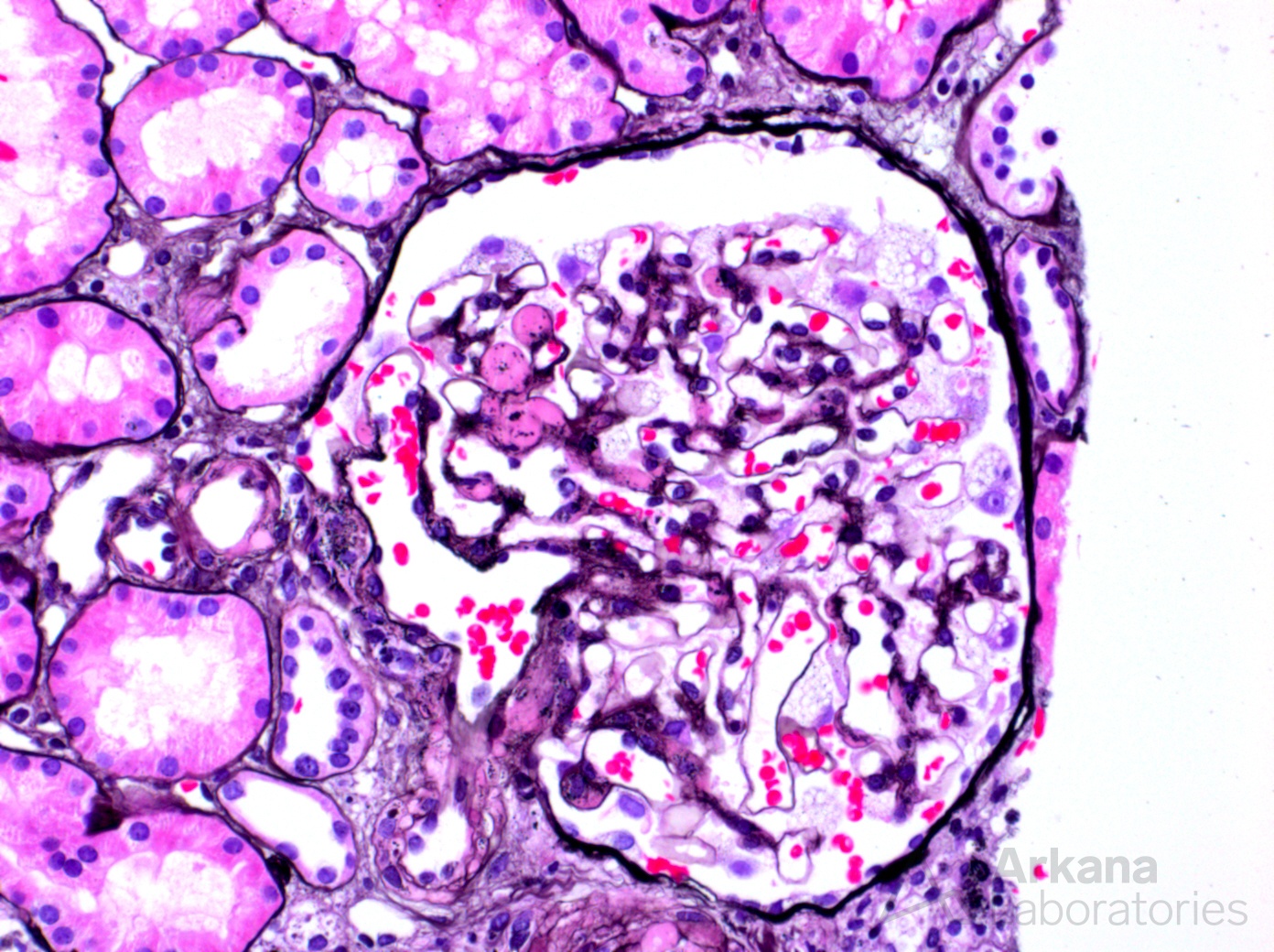
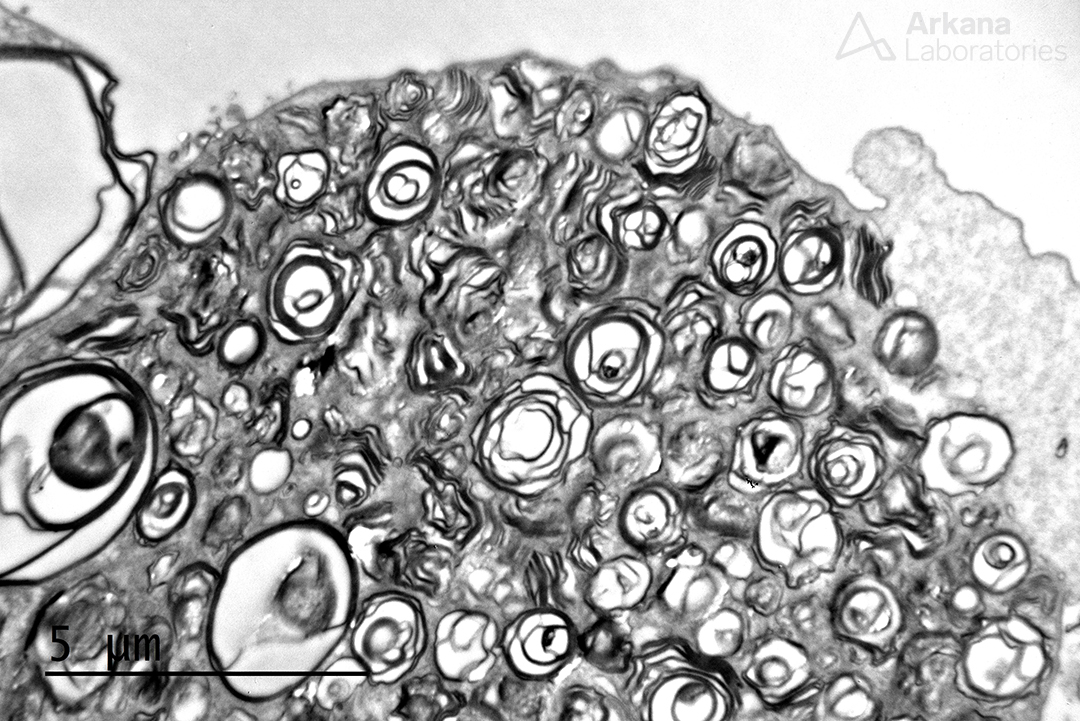
This photomicrograph of a Jones silver stain is interesting in that it shows two findings. First, at 10 o’clock an area of hyalinosis at the perihilar region is present and compatible with perihilar focal segmental glomerulosclerosis. However, if you look closely you will notice that the podocyte cell bodies are very large and vacuolated. This finding is uncommon and is typically seen in the setting of Fabry disease (and usually very difficult to appreciate, unlike in this example). And, while myeloid bodies can be seen in patients on chloroquine or hydroxychloroquine therapy, they are typically sparse or focal in these settings. As suspected, extensive accumulation of intra-podocyte myeloid bodies (a.k.a. zebra bodies, myelinosomes) are seen in all podocyte cell bodies examined (see image below). And, subsequent testing revealed Fabry disease in this patient.
Interestingly, the perihilar focal segmental glomerulosclerosis identified here was present in numerous glomeruli as well as marked glomerulomegaly. While this patient was not obese nor had premature delivery at birth, the biopsy showed around 70% global sclerosis and a second diagnosis of hyperfiltration injury was rendered, likely due to acquired reduced nephron mass secondary to glomerular/nephron loss from the underlying Fabry disease.
Quick note: This post is to be used for informational purposes only and does not constitute medical or health advice. Each person should consult their own doctor with respect to matters referenced. Arkana Laboratories assumes no liability for actions taken in reliance upon the information contained herein.